Introduction to Digital Dentistry
Digital dentistry uses advanced imaging, scanning, and computer-aided design to plan and deliver dental care with greater precision and predictability. Instead of relying only on traditional impressions and hand-made models, we capture data directly from your mouth and transform it into a 3D plan that guides treatment and fabrication. For patients, this often means fewer appointments, improved comfort during records, and restorations that fit more accurately.
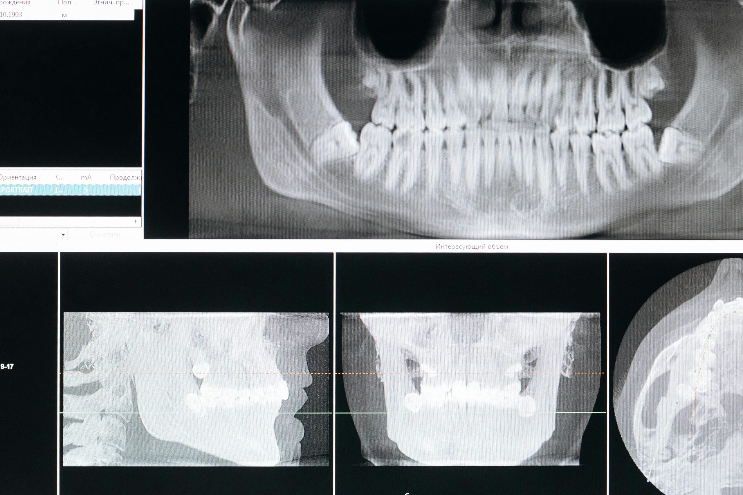
At its core, a digital workflow has three steps: data capture, design, and fabrication. Data capture may include intraoral scanning of teeth and gums, photogrammetry to pinpoint implant positions, and, when appropriate, 3D radiography to visualize bone. Those datasets are aligned in design software to plan the bite, contours, and esthetics. The final design is then transferred to milling or 3D printing to produce provisional and definitive prostheses with high fidelity to the plan.
Full-arch treatment brings specific challenges because small errors can accumulate across a long span. Accurate implant location, stable bite records, and consistent soft-tissue detail all matter, and different tools excel at different parts of that job. Photogrammetry focuses on the exact spatial relationship of implants using reference targets, while intraoral scanners capture soft tissue, teeth, and occlusion. Understanding how each technology contributes sets the stage for comparing photogrammetry vs intraoral scanner full arch workflows and why a carefully coordinated approach can improve outcomes.
From a patient standpoint, preparation is simple and low risk. Maintain your routine oral hygiene with a soft brush; if you wear a removable provisional or denture, rinse it after meals and handle clasps gently to avoid distortion. If you have nightguards, old dentures, or previous models, bringing them to records appointments can help align your new digital plan with how you currently function. Avoid home “DIY impression” kits or attempting to adjust any prosthesis yourself; even minor changes can alter the bite or tissue support in ways that complicate the digital design. If you notice irritation, a sore spot, or a sudden change in how your teeth meet, note when it happens and what you were eating or doing—those details help us verify and refine the digital record during your next visit.
Understanding Photogrammetry in Dentistry
Photogrammetry in dentistry is a camera-based method that calculates the precise 3D position and angulation of dental implants by photographing coded markers from multiple angles. Rather than recording surface textures, it measures how implants relate to one another, independent of soft‑tissue shape or scanner pressure. This is especially helpful when many implants must connect to a single, rigid full‑arch framework.
During capture, small, uniquely coded scan bodies are temporarily attached to the implants. A calibrated camera takes a series of images; software triangulates the codes to solve each implant’s coordinates and orientation within one shared coordinate system. The result is a digital implant map that can be exported to design software and merged with an intraoral scan for soft tissues and bite, or with 3D radiography when indicated.
In comparing photogrammetry vs intraoral scanner full arch records, think of photogrammetry as the skeleton (precise implant axes) and the intraoral scanner as the skin (gums, teeth, and occlusion). Because photogrammetry does not rely on stitching long spans of surface data, it avoids the cumulative drift that can occur over an edentulous arch. Aligning the two datasets lets the lab fabricate a framework that seats passively while honoring the planned contours and bite.
What should you expect? You may be asked to stay still and gently part the lips so all codes are visible; the process is quick and uses photographs, not radiation. Accuracy depends on fully seated scan bodies, proper calibration, and clear line‑of‑sight to each marker; if a code is blocked, additional angles are captured. Photogrammetry records implant positions only—it does not capture soft‑tissue thickness, smile line, or chewing contacts—so it is paired with an intraoral scan or conventional records to complete the picture. For single‑tooth cases, an intraoral scan alone is often sufficient; for full‑arch or immediate conversions, photogrammetry helps minimize fit adjustments and mid‑appointment remakes.
Overview of Intraoral Scanners
Intraoral scanners are handheld optical devices that record the 3D shape of teeth, gums, and how the jaws meet. The result is a precise digital model used to plan care and design restorations or appliances. Scanning is contactless and relies on light, not X‑rays.
Most systems project patterned light and capture rapid image frames; software aligns overlapping frames to build a true‑scale surface mesh. The operator follows a defined path to maintain continuous overlap and coverage, then adds a brief side‑bite scan to relate upper and lower arches. Color capture can highlight margins, tissue biotype, and wear facets, improving communication with the lab. Files export in common formats suited for CAD/CAM design, milling, or 3D printing.
Accuracy is influenced by tissue access and geometry. Stable landmarks (cusps, fossae, palatal rugae), controlled saliva, and gentle retraction help the stitching algorithm stay locked onto the arch. Highly reflective metals and pooled saliva can introduce noise; angling the tip, light air drying, or a thin contrast coating—when appropriate—improves signal. For long, low‑feature spans, clinicians may scan in segments and verify against printed models or intraoral index checks to confirm fidelity.
In implant dentistry, the scanner captures soft‑tissue contours, the emergence profile, and the geometry of scan bodies that encode the implant connection in the CAD design. It is often paired with additional records and verification steps to ensure passive fit before a rigid full‑arch prosthesis is fabricated. Framed within photogrammetry vs intraoral scanner full arch decisions, intraoral scanners supply the detailed surface and bite data, while the overall protocol is adapted to the case to minimize compounding error. At home, if you are prone to gagging, practicing slow nasal breathing can make scanning more comfortable, and avoiding heavily pigmented lip products on the day of records prevents color transfer. If you notice a chip or tenderness around a provisional, pause using that area for tough foods and alert us so we can assess it promptly in person.
Comparing Accuracy in Full-Arch Results
For full-arch frameworks, accuracy hinges on two questions: how closely the record matches reality (trueness) and how consistently it can be repeated (precision). Photogrammetry provides highly consistent implant coordinates across the arch, while intraoral scanning supplies the soft-tissue contours and bite. Used together, they reduce distortion risks that can accumulate over long spans and help achieve a passive, strain-free fit.
Clinically, a “passive fit” means the prosthesis seats without forcing implants toward each other. We verify this with tests such as seating a framework using a single screw at one end and checking for lift at the opposite end, alternating screw sequences to feel for tension, and confirming complete seating radiographically. When the combined records are accurate, screws engage without resistance, the framework does not rock, and occlusion refines with minimal adjustment.
Each method has characteristic error patterns. Photogrammetry errors typically arise from an incompletely seated scan body, obscured markers, or a camera that is out of calibration; these show up as uniform misalignment between multiple implant positions. Intraoral scanner errors are more likely when long, low-feature spans must be “stitched” and when the bite record is captured with inadequate interarch stability; these present as subtle cross-arch twist or an occlusion that is correct on one side and drifts on the other. Merging the two datasets introduces a third checkpoint: the alignment step itself. Anchoring the merge on rigid references tied to the implants, rather than on compressible soft tissues, reduces registration drift and improves the odds of a passive framework.
Timing also matters. During immediate conversion on surgery day, tissue contours are changing, so the implant coordinate map becomes the primary reference; the surface and bite data serve to shape the provisional and are refined as healing progresses. For definitive prostheses, we often add a verification step—such as a printed try-in or a rigid verification jig—to confirm that the digital records behave like the mouth before committing to the final framework. In that context, comparing photogrammetry vs intraoral scanner full arch workflows is less about picking a winner and more about assigning each tool to the part of the job it measures best.
If your full-arch provisional begins to click, rock, or a screw feels loose, avoid repeatedly removing and reinserting it, choose softer foods, and keep the area clean with gentle brushing or a non-alcohol rinse. Please contact us for prompt in-person evaluation during business hours (Mon–Thu 9:00–5:00, Fri 9:00–2:00).
Workflow Differences between Technologies
These approaches diverge most in the order of records, what each record anchors, and how the datasets are verified. A photogrammetry‑led workflow establishes a rigid implant coordinate map first, then adds intraoral scans for soft tissues and bite. An intraoral‑scanner‑only workflow relies on scanning the geometry of implant scan bodies, with additional segmenting and verification steps to control cross‑arch stitching. Both can reach a definitive prosthesis, but the checkpoints and chairside sequence differ.
On an immediate conversion day, photogrammetry is typically performed as soon as the implants (or multi‑unit abutments) are placed and coded markers can be viewed clearly. Once the coordinate set is captured, the markers are removed and an intraoral scan records the arch surfaces, opposing arch, and the bite—often using a prefabricated or existing prosthesis as a jaw‑relation index. The lab merges the datasets to design a provisional that references the fixed implant map. By contrast, an intraoral‑scanner‑only approach captures scan bodies in segments with robust overlap, then validates the digital record with a rigid jig or printed check before fabricating the conversion prosthesis.
For definitive full‑arch work after healing, photogrammetry serves as a coordinate verification while the intraoral scan supplies refined emergence profiles and occlusion at the established vertical dimension. Jaw relation is recorded with a stabilized prosthetic index or bite platform and scanned in position to lock the arches together in the design space. If the case is partially dentate, some teams scan teeth first to “lock” the surface model, then add a brief photogrammetry capture for implant coordinates; in fully edentulous arches, the sequence is commonly reversed so the rigid implant map guides all subsequent records.
Orientation and esthetics are handled differently as well. Some workflows supplement the records with a facial reference or scanned bite fork to maintain the planned incisal plane during design. Where intraoral‑scanner‑only protocols may add a printed try‑in or PMMA prototype to confirm the digital bite and cross‑arch fidelity, photogrammetry‑based protocols often use that try‑in primarily to refine soft tissue and occlusion, since the implant relationship is already locked. In practice, comparing photogrammetry vs intraoral scanner full arch workflows is about choosing which record becomes the master reference and building the rest of the steps around it to minimize compounding error.
Impact on Prosthetic Fabrication
The capture method influences how a full-arch prosthesis is engineered, verified, and manufactured. Photogrammetry supports tighter tolerances for the metal or zirconia framework because implant axes and spacing are locked with high certainty, while intraoral scanning contributes the soft-tissue contours and bite needed to shape the intaglio and occlusion. In practice, this affects whether a framework can be fabricated as a single rigid piece with minimal relief or whether the lab adds compensations, segments the framework, or plans additional verification steps before finish.
On the CAM side, a reliable implant coordinate map allows the lab to nest a one-piece framework, use narrower screw-channel clearances, and target smaller seating allowances at the interface. When the implant geometry comes from scan-body stitching alone, the lab may choose slightly wider channel diameters, increase intaglio relief, or split the framework into sections that are luted extraorally to control cross-arch distortion. The chosen record also guides material strategy: printed provisionals can reference the coordinate map for immediate seating, while definitive frameworks are typically 5‑axis milled with toolpaths and sintering offsets calibrated to the expected tolerance stack.
Design details flow from the soft-tissue and bite data in the intraoral scan. Emergence profiles, flange thickness, and pontic contact are contoured to support hygiene and speech, then harmonized with the occlusal scheme established in the record. When implant positions are resolved by photogrammetry, the lab can prioritize esthetic layering or tooth setup knowing the bar or monolithic substructure will seat passively; functional refinements tend to occur in the teeth rather than at the implant interface. In short, comparing photogrammetry vs intraoral scanner full arch records is less about which file is “better” and more about which dimension (rigid implant fit versus soft-tissue and occlusion) can be trusted to carry the tightest tolerance in fabrication.
At home, if a new bridge has a sharp spot or edge, placing a small piece of orthodontic relief wax over the area is a safe temporary measure until it can be smoothed in person. Clean under fixed spans daily with a floss threader or a water flosser on a low setting aimed sideways along the prosthesis—not directly into the tissue. Do not use adhesives, over‑the‑counter reliners, or attempt to tighten any screws. If you notice rubbing, a chipped tooth surface, or a change in how your teeth meet, please seek prompt in‑person evaluation.
Evaluating Patient Experience and Comfort
Most patients find both record types comfortable because they are light-based and noninvasive. Photogrammetry feels like brief photo taking after small coded pieces are attached to the implants, while intraoral scanning feels like a smooth wand moving along the teeth and gums. For full-arch care, both may be used in the same visit; comfort comes down to how long you need to keep your mouth open, how easily you tolerate retraction, and the ability to pause for short breaks.
With photogrammetry, the main sensations are placement and removal of the coded markers and being asked to hold still while a camera captures multiple angles. There is no vibration or water spray, and captures are typically quick. Because it relies on a clear line of sight, your cheeks and lips may be gently retracted, but the camera itself does not enter deep into the mouth. Patients who are sensitive to gagging often prefer this portion, as it avoids long sweeps near the soft palate.
Intraoral scanning involves a handheld wand that glides across the arches; you may notice gentle retraction, brief air-drying, and the sound of suction. The operator will guide you to rest your tongue away from the scanner and may ask you to swallow between segments to clear saliva. Posterior areas can be the most stimulating; planning short, predictable pauses reduces jaw fatigue. If you have limited opening, the scan can be done in segments so you are never holding a wide stretch for long.
When comparing photogrammetry vs intraoral scanner full arch appointments from the patient’s chair, chair time is usually driven by the surface scan and bite records, while the photogrammetry capture provides a concise “implant map.” You may be asked to gently close into a light index for a few seconds so the software can relate the upper and lower arches. Good communication helps: agree on a hand signal to request a pause, and let us know if a corner of the mouth feels sore so retraction can be adjusted.
At home after a longer records visit, a warm compress to the cheeks can ease jaw tightness, and a lukewarm saltwater rinse is a simple way to freshen tissues. Staying hydrated beforehand and the day of your visit can also reduce dryness during scanning. If any soreness lingers or your bite feels noticeably different afterward, please seek prompt in-person evaluation.
Time Efficiency in Scanning Techniques
Time efficiency in full-arch records is best judged across the whole appointment—setup, capture, and any rework—rather than the stopwatch on a single scan. Workflows that lock a reliable implant reference early tend to reduce pauses and retakes, while surface and bite records determine how many additional passes are needed. In practice, the fastest approach is the one that prevents rescans and minimizes downstream adjustments, not necessarily the one with the quickest individual capture.
Photogrammetry introduces a short setup phase to attach coded markers and confirm camera calibration, followed by rapid image bursts that do not require sweeping through the mouth. Because it solves implant relationships mathematically, the time risk is less about coverage and more about line‑of‑sight; if a code is hidden, a few extra angles are added rather than repeating long spans. Intraoral scanning, by contrast, concentrates time in continuous surface acquisition and the jaw‑relation pass; interruptions that break continuity can add minutes if segments need to be re‑established. Processing time also differs: photogrammetry solves coordinates almost instantly, whereas dense surface meshes may render for a few moments before verification and export.
- Case condition: healed, firm tissues with clear landmarks usually scan faster than mobile or freshly altered tissues.
- Arch features: highly repetitive, low‑feature spans take longer to verify than arches with distinct contours.
- Operator choreography: staging tasks (e.g., assistant exporting files while clinical steps continue) trims idle time.
- Verification strategy: a quick in‑mouth check or printed index can avert longer rescans later.
- Data merge plan: anchoring the alignment on rigid references streamlines integration and reduces back‑and‑forth edits.
On immediate conversion days, decoupling tasks improves throughput: once implant coordinates are solved, other steps—soft‑tissue capture, opposing arch, and bite—can proceed without revisiting implant relationships. For definitive visits, efficiencies come from stable jaw‑relation records and clear emergence profiles so the designer does not request additional captures. In short, when comparing photogrammetry vs intraoral scanner full arch workflows, the time advantage usually comes from how the pieces are sequenced and verified: a brief, dependable implant map paired with a well‑planned surface and bite record avoids the most time‑consuming event of all—a do‑over.
Cost Considerations for Practices
Total cost is more than the sticker on a device; it includes training time, maintenance, accessories, software, and the downstream impact on remakes and chair time. Most practices already invest in an intraoral scanner for broad clinical use. Photogrammetry is typically an additional, case‑selective tool aimed at full‑arch implants where a rigid, passive fit has the highest stakes. The most economical pathway depends on your case mix, available team skills, and how often you deliver immediate conversions or definitive full‑arch frameworks.
Acquisition strategy is driven by utilization. If your workload is primarily single units and short-span restorations, an intraoral scanner may cover the majority of needs. Teams that routinely place or restore multiple implants per arch are more likely to recoup a photogrammetry camera through fewer adjustment visits and reduced risk of framework rework. Practices with low full‑arch volume sometimes collaborate with a lab or a nearby provider who can capture implant coordinates as needed, avoiding underused equipment while maintaining access to the data type.
Ongoing expenses differ between systems. Intraoral scanners carry periodic software updates, calibration tools, and tip sterilization or replacement. Photogrammetry introduces coded markers/scan bodies, a calibration artifact, and instrument servicing; components must remain dimensionally stable, so wear and sterilization protocols matter. File storage, backup, and secure transfer also add predictable, modest costs regardless of modality. When weighing photogrammetry vs intraoral scanner full arch investments, factor the consumable stream and the lifespan of components alongside the capital purchase.
Training and workflow refinement are meaningful cost centers. Establishing calibration routines, capture checklists, and merge protocols takes focused team time. A short learning curve may temporarily slow appointments; the payoff is fewer rescans, fewer verification appointments, and less chairside adjustment during delivery. Minutes saved at delivery often outweigh minutes added to records, especially for rigid, full‑arch prostheses where rework is expensive in both lab and clinical time.
Laboratory relationships influence the balance sheet. Some labs build verification jigs or printed try‑ins by default when records come from scan‑body stitching alone; these steps add shipping and appointment time but serve as insurance against misfit. When reliable implant coordinates are provided, labs may streamline those checkpoints and allocate effort to esthetics and occlusion instead. Clarifying what your lab expects for full‑arch cases—file formats, libraries, and verification preferences—helps avoid duplicate steps and keeps both clinical and lab costs predictable.
Future Trends in Digital Scanning
Digital scanning is moving toward smarter, more connected systems that measure more and guess less. Expect tighter integration between capture devices, automated quality checks during scanning, and standardized data that flows cleanly from clinic to lab. For full‑arch care, the goal is faster, more reliable records that translate into prostheses requiring fewer adjustments.
On the capture side, hybrid approaches are emerging: intraoral scanners that can also resolve implant coordinates with photogrammetric methods, or markerless algorithms that calculate implant pose from known geometries without coded targets. Real‑time “confidence maps” are beginning to flag areas of drift, prompting the operator to rescan a small region before moving on. Dynamic reference frames and improved motion compensation aim to keep long spans stable even if the patient needs short breaks. As the discussion around photogrammetry vs intraoral scanner full arch workflows evolves, you will see fewer either/or choices and more single appointments that gather rigid implant data, soft‑tissue detail, bite, and face orientation in one coordinated session.
Design and verification tools are also advancing. Virtual articulators are being calibrated with jaw‑motion tracking so the occlusion can be tested in simulation before anything is milled. Facial scans—and in some cases light, noncontact 3D photos—will help maintain midline and smile plane without extra physical devices. Automated checksums and calibration logs are likely to accompany scan files, giving labs metrology‑style traceability from capture to CAM. Cloud collaboration with version control will make it easier to compare provisional to definitive records, creating a “digital twin” that documents tissue changes over time and guides where refinement is needed rather than starting over.
For patients, these trends point to shorter visits, fewer try‑ins, and restorations that behave more like the plan because potential errors are caught earlier. The fundamentals remain the same: accurate implant relationships, stable bite records, and clean soft‑tissue detail. If you have a history of jaw fatigue or limited opening, share that ahead of time so we can pace scanning and plan brief pauses. And if anything about your bite or a provisional feels different after records, let us know promptly so we can evaluate it in person.
Conclusion: Choosing the Right Approach
The right approach is the one that reliably delivers a passive, comfortable full‑arch prosthesis with the fewest surprises. In many cases, a blended workflow—photogrammetry for rigid implant coordinates plus an intraoral scan for soft tissue and the bite—offers the strongest foundation. An intraoral‑scanner‑only path can succeed when landmarks are stable and robust verification is planned, while photogrammetry adds confidence when multiple implants span an arch, tissues are changing, or immediate conversion is planned. In short, match the tool to the part of the job it measures best.
Practical selection comes down to case demands and constraints you can’t change on the day: patient anatomy (limited opening, cheek bulk), visibility to implant markers, the need for a rigid one‑piece framework, and the team’s scanning proficiency and lab coordination. If line‑of‑sight to coded markers will be difficult, plan retraction and capture angles in advance or stage the scan; if long, low‑feature spans are expected, structure the intraoral scan in segments with deliberate overlap and a stable jaw‑relation index. Agree with your lab on libraries, file formats, and how the datasets will be merged before the appointment so the “master reference” is clear.
Risk management is about tolerances and checkpoints. Define what must be exact (implant fit), what can be adjusted (occlusion and contours), and where you will verify before committing to a definitive framework. When uncertainty arises—ambiguous merge, limited visibility, or motion during capture—choose the conservative detour: a printed try‑in, a rigid verification jig, or a brief re‑capture that targets the weak link rather than repeating everything. Framed this way, the photogrammetry vs intraoral scanner full arch decision is simply a plan for where accuracy is anchored and how you will prove it.
For patients, the takeaway is straightforward: expect light‑based records that are generally quick and comfortable, with pauses for rest as needed. After longer visits, a small amount of bland lip balm can soothe the corners of the mouth if they feel dry from retraction. If a provisional feels loose, begins to click, or your bite feels different, avoid home fixes and seek prompt in‑person evaluation so we can check the records and the prosthesis together. Thoughtful planning and the right capture sequence keep your treatment on track and reduce the need for adjustments later.
Frequently Asked Questions
Here are quick answers to common questions people have about Photogrammetry vs IOS for Full-Arch Accuracy in Glendale, AZ.
- What is the main advantage of using photogrammetry in full-arch dental procedures?
Photogrammetry offers high precision in capturing the exact 3D position and angulation of dental implants. It avoids cumulative errors that can occur with long spans and provides a reliable implant coordinate map crucial for fabricating a rigid full-arch framework. This technology focuses on the ‘skeleton’ of implant relationships, making sure they align correctly without being influenced by soft-tissue shapes or pressure.
- How does an intraoral scanner contribute to full-arch dental workflows?
Intraoral scanners provide a detailed digital model of the teeth, gums, and occlusion. They are crucial for capturing soft tissue contours, the emergence profile, and the geometry of scan bodies in implant dentistry. This scanner acts as the ‘skin,’ supplying valuable surface data needed for designing restorations and ultimately ensuring a fit that considers both comfort and esthetic detail.
- Why are both photogrammetry and intraoral scanning used together in full-arch treatments?
Using both technologies together combines the precision of photogrammetry for implant positioning with the comprehensive soft-tissue and bite data from intraoral scanners. This reduces the risk of fit distortion over long spans and ensures a passive, strain-free fit for prostheses, which is critical for avoiding adjustments and ensuring long-term success.
- What should patients expect during a full-arch appointment involving photogrammetry and intraoral scanning?
Patients can expect a non-invasive and comfortable procedure, where photogrammetry involves brief photo capture of implants using coded markers, and intraoral scanning includes the gentle movement of a wand across the teeth and gums. The entire process is light-based and might require some retraction but generally allows for breaks and communication to ensure comfort.
- How do photogrammetry and intraoral scanners differ in terms of workflow timing?
Photogrammetry involves a brief setup to attach coded markers and capture implant data quickly with minimal need for retakes. Intraoral scanning focuses on continuously capturing surface and bite data, which takes time to ensure complete coverage. The most time-efficient workflow is the one that minimizes the need for rescans and reduces the chance of downstream adjustments.
- What cost considerations should practices evaluate when choosing between photogrammetry and intraoral scanning?
Practices need to consider not only the purchase cost of the devices but also expenses related to training, maintenance, and workflow refinement. Photogrammetry may be more cost-effective for practices handling multiple full-arch implant cases, potentially reducing the need for adjustments and remakes. Intraoral scanners are versatile and widely used, often forming the foundation of digital workflow in dental practices.
Medical sources (PubMed)
- Al-Nawas B, et al. Int J Implant Dent. 2023. “ITI consensus report on zygomatic implants: indications, evaluation of surgical techniques and long-term treatment outcomes.”. PMID: 37698775 / DOI: 10.1186/s40729-023-00489-9
- Schwarz F, et al. Periodontol 2000. 2022. “It is all about peri-implant tissue health.”. PMID: 35103327 / DOI: 10.1111/prd.12407
- Sanz-Sánchez I, et al. Periodontol 2000. 2022. “Complications in bone-grafting procedures: Classification and management.”. PMID: 35103322 / DOI: 10.1111/prd.12413
- Monje A, et al. Periodontol 2000. 2022. “Management and sequelae of dental implant removal.”. PMID: 35103326 / DOI: 10.1111/prd.12418
- Michelinakis G, et al. BMC Oral Health. 2021. “The direct digital workflow in fixed implant prosthodontics: a narrative review.”. PMID: 33478459 / DOI: 10.1186/s12903-021-01398-2
- Chackartchi T, et al. Periodontol 2000. 2019. “Soft tissue-related complications and management around dental implants.”. PMID: 31407443 / DOI: 10.1111/prd.12287