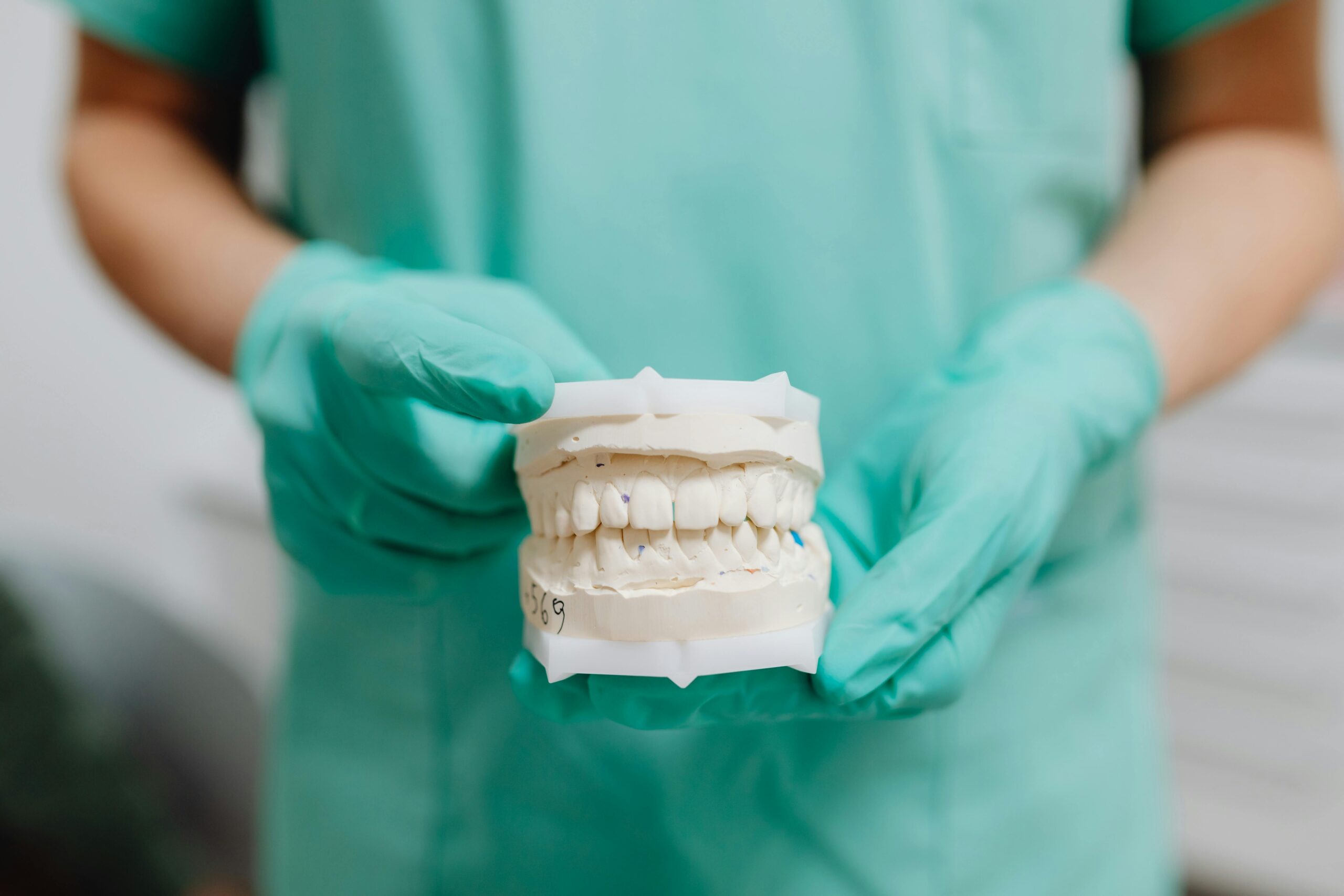
Understanding Endodontic File Separation
Endodontic file separation means a small piece of a root canal instrument has broken inside the tooth during treatment. It happens because these thin tools work in curved, narrow canals under stress. While unsettling to hear, a separated file does not automatically mean treatment failure; many cases can still heal well with careful planning. Your dentist or endodontist will explain what it means for your specific tooth and outline safe options.
Files typically fracture in two ways: torsional failure (the tip binds while the shank keeps turning) and cyclic fatigue (repeated bending in a curve), with risk increasing in tight, curved, or calcified canals and with instrument reuse [1]. Understanding how canals are shaped and cleaned helps put this into context; see our concise overview of what happens during root canal treatment.
- Where the fragment sits (near the tip versus higher in the canal)
- Canal curvature and width, and whether the piece blocks cleaning
- Signs of infection before or during treatment
- Instrument type and size, and how long it was used
Management usually starts with imaging and magnified inspection. If possible, the clinician may “bypass” the fragment with small files to clean around it, or attempt retrieval using ultrasonics and microtubes under a microscope; success depends on location, canal anatomy, and dentin preservation [2]. When the fragment is very deep or past a sharp curve, it may be safer to leave it, complete disinfection as far as possible, and monitor the tooth over time. Referral to an endodontist is common when specialized tools or experience are needed.
Most approaches follow practical endodontic file separation guidelines: communicate clearly with the patient, assess risks and benefits of retrieval versus retention, prioritize infection control, and document findings and decisions. The goal is the same in every case—achieve the best long-term health for the tooth while minimizing unnecessary structural loss. If you have questions about how this applies to your situation, ask your clinician to walk you through the plan step by step.
Importance of Following AAE Guidelines
Following the American Association of Endodontists (AAE) guidance helps clinicians respond to a separated file in a calm, structured way. Clear steps—informing you, assessing the tooth, and choosing whether to remove or work around the fragment—reduce risk and support tooth survival. These recommendations also promote careful documentation and follow-up, so everyone understands the plan and expected outcomes. Put simply, AAE guidance turns a stressful moment into a safer, more predictable process.
AAE-aligned care emphasizes informed consent, meaning your dentist explains what happened, why it happened, and the options. It also stresses thorough evaluation: using magnification, appropriate imaging, and gentle techniques that protect dentin. Rather than chasing every fragment, the focus is on infection control and preserving tooth structure, because over-cutting can weaken the tooth more than the fragment itself. By following endodontic file separation guidelines, the team balances benefits and risks at each step and keeps your long-term tooth health at the center.
Another key point is knowing when to refer. If the fragment sits deep in a curve or removal would remove too much tooth, an endodontist with specialized microscopes and micro-instruments may be best suited to help. If retrieval is not advisable, disinfecting around the fragment and sealing the canal can still lead to good healing in many cases. Planned follow-up—checking symptoms and periodic imaging—helps confirm that the tooth stays comfortable and the bone around the root remains healthy over time.
For you as a patient, good guidelines mean clear communication and a thoughtful plan: what will be done today, what risks are reasonable, what signs to watch for, and how the tooth will be protected afterward. Ask how the clinician will minimize extra drilling, whether referral is recommended, and how the restoration will strengthen the tooth once treatment is complete. In many cases, that includes discussing how a protective crown is planned after root canal therapy to reduce future fracture risk. The goal is steady, evidence-informed care that protects both your comfort and your tooth.
When to Leave a Separated File
It is generally appropriate to leave a separated file in place when trying to remove it would require cutting away too much dentin, risk a perforation, or push the fragment deeper. Leaving the fragment is also reasonable when it lies deep in the apical third or past a sharp curve, cannot be safely bypassed, and you can still disinfect and seal the canal effectively. If the tooth was not showing signs of active infection and the fragment does not block cleaning, the chance of healing can remain good. In all cases, the choice follows a careful, individualized risk–benefit discussion.
Your clinician first assesses where the fragment sits (coronal, mid, or apical third), canal curvature, and whether it blocks working length. Magnification and imaging help determine if the piece can be bypassed or if limited ultrasonic exposure could uncover it safely. If a fragment prevents adequate cleaning in a tooth with active infection, removal or alternative access (including referral) is weighed more strongly. When cleaning around the fragment is achievable and a tight coronal seal can be placed, retention with monitoring is often the safer path.
Tooth preservation drives the decision. Aggressive attempts can create ledges, cracks, or strip perforations that compromise long-term strength more than the fragment itself. That is why endodontists use microscopes, conservative ultrasonics, and “dentin-sparing” strategies—and why they may recommend leaving the fragment when the anatomy is unfavorable. If a retrieval or extended procedure is planned, discuss comfort options such as gentle oral sedation for complex endodontic visits.
Follow-up is essential. Your dentist will document the event, complete disinfection as far as safely possible, and seal the tooth well to block new bacteria. Expect periodic reviews—symptom checks and imaging—to confirm quiet tissues and stable bone. Report new pain, swelling, or a gum pimple (sinus tract) promptly, as these may signal the need for further care such as surgical access or retreatment. These choices align with practical endodontic file separation guidelines: protect dentin, control infection, and monitor results over time. If you are planning a visit or have timing questions, you can check our current hours.
Bypassing a Separated Endodontic File
Bypassing means gently sliding a very small hand file around a broken piece so the dentist can reach full length again and clean the canal. It is often the first choice when the fragment is visible or sits before a sharp curve, because it preserves tooth structure and avoids aggressive cutting. If a pathway is gained, the canal can be disinfected and shaped around the fragment so irrigants can reach the tip of the root.
How is it done? Under a dental microscope, the clinician creates a tiny, smooth platform and uses pre-curved stainless-steel files (usually sizes 6–10) with light, patient movements. Lubricants and frequent irrigation help the file “find” a track alongside the fragment without pushing it deeper. An electronic apex locator confirms length, and the path is gently widened to allow irrigants and medication to flow past the obstruction. If the fragment can be safely loosened, it may be removed; if not, a stable bypass with good cleaning can still lead to healing.
When will bypassing work? Odds are better when the fragment is higher in the canal, above or at the start of a curve, and when there is space to work under magnification. Deep apical fragments, severe curves, or very tight canals lower the chance and raise the risk of unnecessary dentin loss. Research has measured how often bypassing succeeds and how long it can take, emphasizing that results vary with instrument design and clinical conditions [3]. Following endodontic file separation guidelines, your dentist weighs these factors and discusses whether to bypass, attempt limited ultrasonic uncovering, or leave the fragment and focus on disinfection.
Thorough cleaning remains the goal. Even if the fragment stays, strong irrigation and activation strategies help carry disinfecting solutions around it to reduce bacteria. This improves the quality of cleaning despite the blockage [4]. Throughout, the team protects dentin, avoids heavy forces that could ledge or perforate, and seals the tooth well at the end of treatment. Planned follow-up checks comfort and healing on images over time, and referral to an endodontist is advised when specialized tools or experience are needed.
Retrieving a Separated File: Best Practices
Retrieval is attempted only when the benefits outweigh the risks to your tooth. The safest approach uses a dental microscope, gentle ultrasonics, and conservative dentin removal to expose and loosen the fragment. Success is most likely when the piece is higher in the canal and in a straighter segment; deeper or sharply curved fragments are harder and riskier. If removal becomes unsafe, the plan shifts to bypassing or leaving the fragment while focusing on thorough disinfection.
First, the tooth is isolated with a rubber dam and carefully re-imaged; a microscope exam confirms visibility and access. The clinician removes minimal dentin to create a small “staging platform,” straightens the path just enough to see the fragment, and verifies working length with an electronic apex locator. Irrigation and lubricants keep debris moving, prevent the fragment from wedging, and cool the area during ultrasonic use.
Ultrasonics are applied in short, light bursts to “unseat” the fragment, often starting counterclockwise to disrupt how it is locked in place. Intermittent activation with ample irrigation controls heat and reduces the chance of cracks. If the fragment starts to spin or migrate coronally, it may be teased out or captured using microtube-and-loop methods. Throughout, the goal is to preserve dentin; removing too much to “chase” a fragment can weaken the root or cause a perforation.
If visualization is poor, a limited cone-beam CT may be considered to map exact position and canal curvature. When retrieval efforts risk pushing the fragment apically, creating ledges, or removing excessive tooth structure, stopping is the safer choice. In that case, the team may bypass the fragment, enhance irrigation and activation to clean around it, place intracanal medication if indicated, and seal the tooth well.
Clear communication, informed consent, and documentation anchor every step and align with endodontic file separation guidelines. Your dentist or endodontist will explain the plan, the likelihood of retrieval, alternatives if it proves unsafe, and the follow-up schedule. The measure of success is not just whether the fragment is removed, but whether the tooth heals and stays strong over time.
Implications of File Separation on Treatment
A separated file changes how your root canal is finished, but it does not always change the final outcome. The key question is whether the canal can still be cleaned and sealed to its full, safe length. If cleaning remains possible, treatment can often be completed as planned and the tooth monitored. If the fragment blocks cleaning, your dentist will discuss options such as bypassing, removal, or a staged approach with referral.
Healing depends on where the fragment sits and whether infection is present. A piece that is higher in the canal and does not block disinfecting usually carries a good outlook. A fragment deep near the tip, especially in a tight curve, can make cleaning harder and may raise the risk of ongoing infection. In those cases, the team weighs the benefits of retrieval against the risk of removing too much dentin, and may choose to work around the fragment or plan a different access if needed.
Expect changes to the workflow. There may be more time under a microscope, additional imaging, and careful use of small files and ultrasonics. Sometimes the tooth is dressed with medication and closed temporarily so progress can be checked at a second visit. Throughout, endodontic file separation guidelines emphasize clear communication, informed consent, and documentation, so you understand what happened, what will be done today, and what to expect next.
Protecting tooth strength is a major consideration. Aggressive attempts to remove a deep fragment can thin the root or cause a ledge or perforation, which can harm long-term success more than the fragment itself. Whatever approach is chosen, a tight coronal seal at the end of treatment helps block new bacteria from entering the canal. Many teeth also benefit from a full-coverage restoration to reduce future fracture risk; your dentist will explain timing and materials based on how much tooth structure remains.
Follow-up matters. Your care team will check comfort, test the tooth, and review periodic images to confirm quiet tissues and healthy bone around the root. Call promptly if you notice new pain, swelling, or a gum pimple (sinus tract), as these signs can signal the need for retreatment or surgical care. The goal is the same in every case: control infection, protect dentin, and help the tooth stay comfortable over time.
Assessing the Clinical Situation
Assessment starts with two big questions: can we still clean and seal the canal safely, and would trying to remove the fragment harm the tooth more than help? Your dentist checks where the piece sits, how curved and narrow the canal is, and whether there are signs of infection. They also consider how much healthy tooth remains and whether a strong final restoration is possible. These points guide whether to retrieve, bypass, or leave the fragment with careful monitoring.
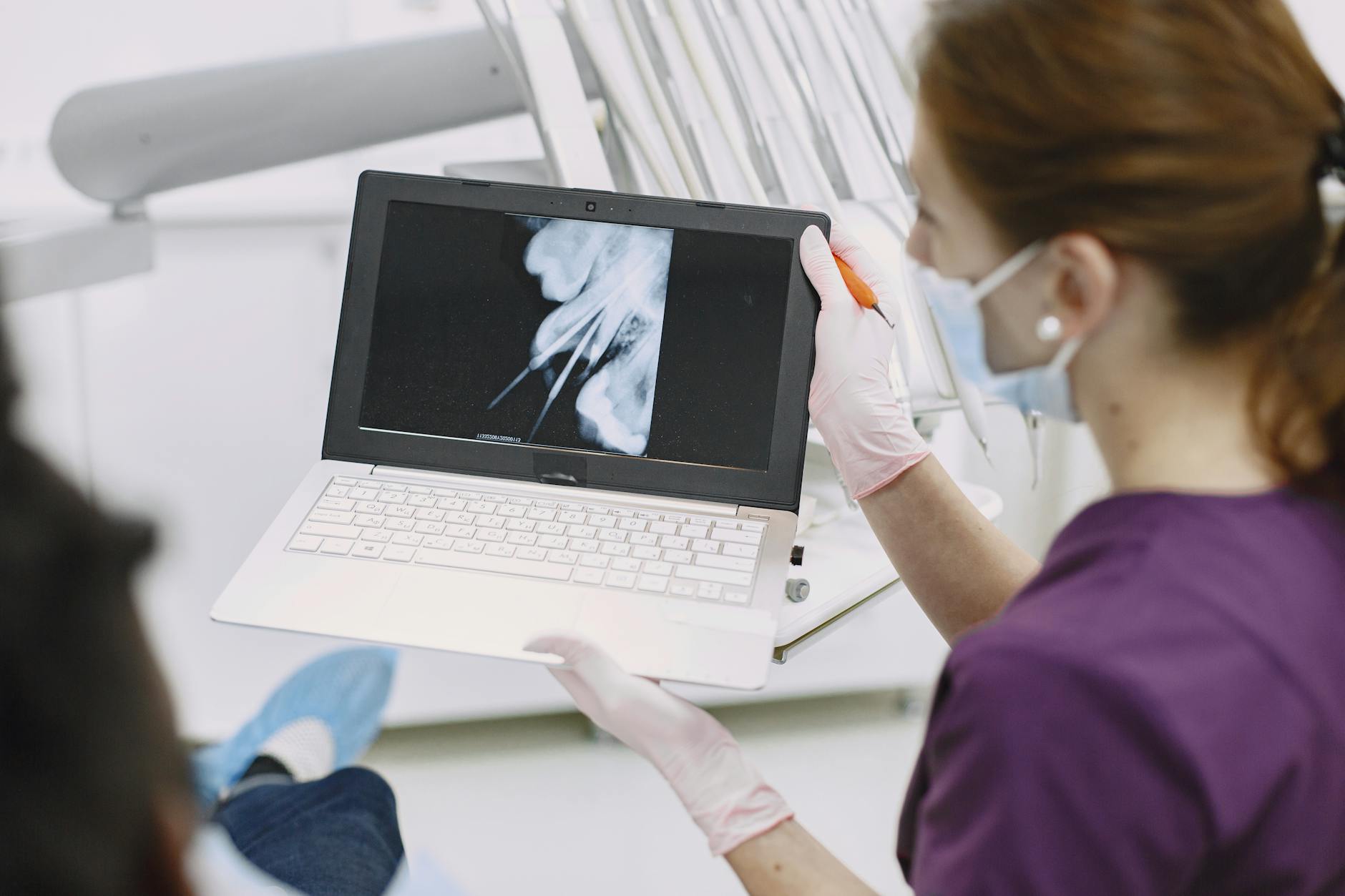
The visit usually begins with a clear history and exam: current pain or swelling, prior symptoms, and any sinus tract. Periapical radiographs from different angles help estimate the fragment’s level (coronal, middle, or apical third) and any bone changes. Under magnification, the clinician verifies working length, tests for patency, and decides if a small hand file can slide past the fragment. If the piece blocks disinfection in an infected tooth, removal or an alternate plan is weighed more strongly; if cleaning around it is feasible, conservation often wins.
Imaging is tailored to need. Extra angled radiographs are routine; a small field CBCT may be considered when three‑dimensional details (exact position, canal curvature, nearby anatomy) would change the plan. The goal is to preserve dentin while controlling bacteria. That means using gentle ultrasonics only when exposure is safe, avoiding heavy forces that could create ledges or perforations, and confirming that a tight coronal seal can be achieved afterward. Endodontic file separation guidelines support this structured approach: prioritize disinfection, weigh risks and benefits, and protect tooth strength.
Patient factors also matter. Strategic value of the tooth, ability to restore it well, medical considerations, and comfort needs are part of the decision. Your dentist will explain options—retrieval, bypass, or leaving the fragment—along with expected benefits, risks, and alternatives, and document the plan and consent. Follow‑up is set to check symptoms and healing on images; new pain, swelling, or a gum pimple should be reported promptly. Referral to an endodontist is recommended when specialized tools or experience can improve safety or outcomes.
Managing Patient Expectations
After a file separates during a root canal, you should expect a calm, clear explanation and a step‑by‑step plan. Most teeth can still heal well; success depends on how safely the canal can be cleaned and sealed. Your dentist will outline choices—remove, work around, or monitor the fragment—and explain what will be done today and what comes next. These conversations follow endodontic file separation guidelines so decisions balance safety, tooth strength, and comfort.
At the visit, we start by listening to your concerns, then review images and show where the fragment sits. We describe why it happened, what it means for your tooth, and which options are reasonable in your case. You will hear the expected time needed, whether a second visit is likely, and if referral to an endodontist would improve safety. We also discuss comfort measures during treatment and how we will protect remaining tooth structure.
We set realistic goals together. “Success” is not only removing the fragment; it is getting the tooth comfortable, controlling infection, and preserving strength. If removal risks too much dentin loss, we may choose to bypass or leave the fragment while focusing on thorough cleaning and a tight seal. You will know what is planned today, what signs to watch for, and when we will reassess.
Expect careful documentation and informed consent. We record what occurred, what was discussed, and why the chosen plan fits your anatomy and symptoms. If a temporary medication is placed, we will explain how to care for the tooth and when the final restoration will be done. Avoid chewing hard foods on the tooth until it is fully protected with its final restoration, as advised by your clinician.
Follow‑up is part of the plan. We check how the tooth feels and review images over time to confirm quiet tissues and healthy bone. Call your dentist promptly if you notice new pain, swelling, or a gum pimple, as these can signal the need for further care. Our aim is steady, evidence‑informed care that reduces worry, protects dentin, and helps your tooth stay healthy over the long term.
Post-Procedure Care for Patients
After a root canal where a file separated, most people feel mild soreness for a day or two, similar to a bruise. This is usually managed with over‑the‑counter pain relievers and gentle care at home. Avoid hard chewing on the treated tooth until your dentist finishes the restoration, because the tooth can be weaker before it is fully protected. Clear instructions, follow‑up, and simple home care are the focus right after treatment.
For comfort, take the pain medicine your dentist recommends. Many patients do well with ibuprofen, acetaminophen, or a combination if medically appropriate; if you cannot take anti‑inflammatory medicines, acetaminophen alone is often used. Some tenderness to biting can be normal at first. A cold compress on the cheek (10 minutes on, 10 minutes off) may help the first day.
Keep the area clean with gentle brushing and your usual mouth rinse; warm salt‑water rinses (½ teaspoon salt in a cup of warm water) can soothe sore gums. If you have a temporary filling, avoid sticky, hard, or very crunchy foods on that side. If the temporary feels loose or comes out, contact your dentist so the tooth can be resealed promptly.
Antibiotics are not routinely needed after endodontic treatment. They are considered when there are signs of spreading infection, such as facial swelling, fever, or you feel generally unwell. Call your dentist promptly if you notice increasing pain after the first few days, swelling, a bad taste that persists, or a pimple on the gum near the tooth—these signs may mean the tooth needs further attention.
Plan for the next steps. Your dentist will complete disinfection as safely as possible around the fragment and place a well‑sealed restoration. Many teeth benefit from a protective full‑coverage restoration to reduce future fracture risk; your dentist will explain timing based on how much natural tooth remains. You will also be scheduled for check‑ins to make sure the tooth stays comfortable and the surrounding bone looks healthy on images.
Throughout, care follows practical endodontic file separation guidelines: communicate clearly, protect tooth structure, control bacteria, and monitor results over time. If anything feels unusual or you are worried, reach out—early communication helps keep you comfortable and on track for healing.
Risks Associated with File Separation
When a file breaks during a root canal, the main risks relate to cleaning and the tooth’s strength. A fragment can block access to the tip of the root, making it harder to remove bacteria and inflamed tissue. Attempts to remove the piece can also require extra drilling, which may weaken the root. Careful planning aims to reduce these risks while still completing safe treatment.
The biggest concern is infection control. If the fragment prevents full cleaning in a tooth that already shows signs of infection, the chance of lingering bacteria is higher. When the fragment sits higher in a straighter part of the canal, the dentist can often bypass or remove it, restoring access. If it lies deep near the tip or beyond a sharp curve, removing it may cause more harm than good. In those cases, the team focuses on disinfecting as far as safely possible and sealing the canal tightly so new bacteria can’t enter.
Procedural risks come from chasing a fragment too aggressively. Creating a wide “staging” area or prolonged ultrasonic use can remove excess dentin, create ledges, or even cause a strip perforation. These complications can reduce long‑term tooth strength and may require additional care. That is why clinicians work under magnification, use light forces, and stop if the removal path becomes unsafe. Preserving dentin is often the safer choice than forcing retrieval at any cost.
Other risks are practical. Treatment can take longer, may need an extra visit, and sometimes requires referral to an endodontist with specialized tools. If symptoms continue despite careful cleaning, the tooth might need retreatment or a surgical approach to reach the tip of the root. Throughout, clear communication and documentation help you understand what happened, why a given choice is recommended, and what signs to watch for during healing.
Following endodontic file separation guidelines helps manage these risks: be transparent, assess whether full cleaning is possible, balance retrieval against dentin preservation, and ensure a strong final seal. The goal is steady, evidence‑informed care that keeps you comfortable and gives the tooth the best chance to heal and stay strong over time.
Evaluating Success After Separation Management
Success means the tooth is comfortable, usable, and shows healthy healing on images over time. Whether the fragment was removed, bypassed, or left in place, the key test is infection control with a strong seal that protects the tooth. Your dentist checks symptoms, function, and X‑rays at set intervals to confirm the plan is working.
Right after treatment, a baseline radiograph is taken and the tooth is sealed well. In the first days, mild tenderness can be normal. Lasting or increasing pain, swelling, or a gum pimple (sinus tract) suggests the tooth needs a closer look. If you can chew gently without sharp pain and the bite feels normal, that is a good early sign.
At follow‑up (often 6–12 months), your dentist reviews three things. First, symptoms: no spontaneous pain, no swelling, and normal responses to gentle tapping and palpation. Second, function: you can chew comfortably and the restoration stays intact. Third, imaging: if there was a pre‑treatment dark area at the root tip, it should shrink or resolve; if none was present, images should remain stable. Bone can take time to fill in, so improvement rather than perfection is often the realistic marker in the first year.
If a fragment was left in place but the canal was disinfected and sealed, many teeth heal well. What matters is that irrigants reached the working length, a clean path was established or closely approximated, and the coronal seal stays tight. If the dark area grows, a sinus tract persists, or symptoms return, your team may consider retreatment, surgical access to the root tip, or referral to an endodontist.
Documentation supports decisions: where the fragment sits, how it was managed (retrieved, bypassed, or retained), the final working length, and the quality of the seal. These points align with endodontic file separation guidelines, which emphasize infection control, dentin preservation, and planned monitoring. Expect another check at 18–24 months if a lesion was present, because some roots show continued healing over a longer window. Throughout, the aim is steady, measurable progress—quiet tissues, stable function, and healthy bone on images.
Summary of AAE Recommendations
The American Association of Endodontists (AAE) recommends a calm, step‑wise approach when a file separates: inform the patient, assess the tooth carefully, and choose the safest path to control infection. Decisions center on whether to retrieve the fragment, bypass it, or leave it in place while maintaining effective cleaning and a strong seal. Throughout, the guidance emphasizes protecting dentin, documenting decisions, and arranging follow‑up.
Initial steps include transparent communication and a focused evaluation under rubber dam isolation. Angled periapical radiographs and magnification (microscope or loupes) help locate the fragment and judge canal curvature; a small‑field CBCT may be considered only if three‑dimensional details would change the plan. Working length and patency are reassessed with gentle, small hand files, avoiding force that could push the fragment apically or create ledges.
The decision framework is risk‑benefit based. Retrieval is considered when the fragment is higher in the canal, visible, and accessible in a relatively straight segment. Bypassing is often preferred when a safe pathway can be made alongside the fragment without removing unnecessary dentin. Leaving the fragment is reasonable when it lies deep in the apical third or beyond a sharp curve, especially if removal would weaken the root or risk perforation, and if the canal can still be disinfected and sealed effectively. Informed consent and thorough documentation accompany each choice, consistent with endodontic file separation guidelines.
Technique recommendations focus on conservation. If retrieval is attempted, a small staging platform and light ultrasonics under high magnification are used, with irrigation to cool and carry debris; efforts stop if dentin removal becomes excessive or visualization is lost. Whether the fragment is removed or retained, irrigation and activation strategies are emphasized to improve disinfection, and a tight coronal seal is placed. Referral to an endodontist is advised when specialized tools or experience would improve safety or outcomes.
Follow‑up is planned to confirm healing and comfort. The AAE encourages monitoring symptoms and periodic imaging (often at 6–12 months, and longer if a lesion was present). Changes such as persistent pain, swelling, or a new sinus tract prompt reassessment and consideration of retreatment or surgical options. The overarching goal remains the same: control bacteria, preserve tooth structure, and protect long‑term tooth function.
Frequently Asked Questions
Here are quick answers to common questions people have about Separated Files in Endo: What the AAE Recommends in Glendale, AZ.
- What is endodontic file separation?
Endodontic file separation happens when a piece of the instrument used during a root canal breaks inside the tooth. This is usually due to the stress of working in tight, curved canals. Though it might sound alarming, a separated file does not necessarily mean that the treatment will fail. Many teeth can still heal successfully with careful planning and management. Your dentist or endodontist will evaluate the situation and discuss safe treatment options tailored to the specific circumstances of your tooth.
- What causes a file to separate during a root canal?
Files can separate in two main ways during a root canal: torsional failure and cyclic fatigue. Torsional failure occurs when the file’s tip gets stuck while the rest of the tool continues to twist. Cyclic fatigue happens due to repeated bending in curved canals. The risk of these issues increases in canals that are particularly tight, curved, or calcified, and when instruments are re-used. Regular assessment and careful operation of the tools can help minimize these risks.
- How is a separated endodontic file managed?
Management often starts with imaging and a close inspection under magnification. Dentists might attempt to bypass the fragment with small files to clean around it or try to retrieve it using ultrasonics and microtubes. Success depends on the file’s location and canal anatomy. Some cases may require a referral to an endodontist if specialized tools or experience are necessary. Treating professionals follow guidelines to balance the risks of retrieval against the benefits of maintaining tooth structure.
- When might a separated file be left in place?
A separated file might be left in place if trying to remove it would cause more harm, like cutting away too much tooth or risking a perforation. The fragment could be retained if it is in a position that allows effective disinfection and sealing of the canal. This could be the safer option if the piece does not block cleaning and the tooth was not previously infected. Retaining the fragment while monitoring can preserve the tooth’s long-term strength.
- Why are AAE guidelines important in managing file separation?
The American Association of Endodontists (AAE) guidelines provide a clear, organized approach that helps manage the situation safely and effectively. By following these recommendations, clinicians can significantly reduce risks, facilitate better communication with patients, and improve the chances of tooth survival. The guidelines focus on infection control, preserving tooth structure, and ensuring that all actions align with the best patient outcomes over time.
References
- [1] Fractographic analysis of separated endodontic file designs. (2020) — PubMed:33140130 / DOI: 10.1007/s10856-020-06432-3
- [2] Management of separated endodontic instruments: case reports. (2022) — PubMed:34978988
- [3] Success Rate and Time for Bypassing the Fractured Segments of Four NiTi Rotary Instruments. (2017) — PubMed:28808464 / DOI: 10.22037/iej.v12i3.16866
- [4] Effectiveness of different irrigation systems in the presence of intracanal-separated file. (2019) — PubMed:30636084 / DOI: 10.1002/jemt.23165