Understanding CBCT Technology
Cone beam computed tomography, or CBCT, is a 3D dental X‑ray. The machine rotates once around your head with a cone‑shaped beam, collecting many small images. A computer then assembles these into a detailed 3D view of teeth, jaws, and nearby structures. Dentists use CBCT when standard 2D X‑rays may not show enough detail.
CBCT works by balancing image quality with exposure. The operator selects a field of view, which limits the scan to the exact area of interest. Smaller fields focus on the problem site, and can reduce exposure while giving fine detail. Settings such as voxel size, kV, mA, and scan time are tailored to the task, not one‑size‑fits‑all. Understanding cbct radiation dose safety starts with these choices and the principle of keeping dose as low as reasonably achievable.
Modern units also use pulsed X‑rays, careful collimation, and motion‑reduction tools. These features send X‑rays only when needed, shape the beam to the target, and help prevent blurring. The result is clear anatomy with less scatter. For example, locating tiny extra canals during complex root canal care often benefits from these settings. When metal is present, software can reduce some streaks, though not all artifacts can be removed.
Clinically, CBCT helps answer specific questions. It is valuable for impacted teeth, implant planning, jaw joints, and sinus relationships. Example: planning the removal of a deeply impacted wisdom tooth. A focused scan can show the tooth’s roots and the nerve canal to guide safer surgery, as in complex wisdom tooth removal planning. CBCT is not routine for every patient; it is chosen when the expected benefit outweighs the small added exposure compared with 2D imaging.
Knowing how CBCT creates images sets the stage for understanding how settings and design features keep exposure appropriate for the task. Next, we look at how dose is minimized and how safety is built in. The right scan, for the right reason, at the right settings.
Radiation Dose Comparisons
CBCT dose varies with scan size and settings, so comparisons are about ranges, not one fixed number. A small, focused CBCT is often similar to, or a bit higher than, a panoramic X‑ray. Larger scans can be higher than a few small 2D images, yet they are far below the dose from a medical CT of the head. The key is matching the scan to the clinical question.
Real‑world example: planning a single implant in a back tooth. A small field of view targets just that area, giving 3D detail with a dose in the same neighborhood as a panoramic image. Medium or large fields, used when both jaws or sinuses must be seen, use more radiation because they cover more anatomy. For full‑arch work, such as planning full arch implant treatment, the larger view can be appropriate because it guides nerve and sinus safety across the entire jaw.
Why do these differences exist? CBCT follows simple physics: more area and higher resolution need more photons. Operators balance three levers, field of view, exposure settings, and voxel size. Smaller fields reduce scatter and dose, lower mA and optimized kV cut output, and slightly larger voxels can still answer many clinical questions. Many units pulse the beam and collimate it to the target, which reduces unnecessary exposure and the chance of repeat scans due to motion. These design choices support cbct radiation dose safety in daily practice.
For patients, the comparison that matters is benefit versus added exposure. If a 2D X‑ray answers the question, it is chosen. If 3D detail will change the plan, shorten surgery, or avoid surprises, a focused CBCT can be the safer overall path. Age, anatomy, and the problem at hand guide the choice, and your dentist can explain why a particular scan size is recommended. Next, we look at how protocols and quality checks keep exposure low while preserving the detail needed for accurate diagnosis and treatment. The right scan, at the right size, for the right reason.
Everyday Exposures Explained
We all receive small amounts of natural radiation every day from the ground, air, and space. A dental CBCT adds to that background, but the amount depends on how large the scan is and how it is set. For many focused scans, the added exposure is small compared with what we all get over time from daily life.
Imagine a two-hour flight to visit family. Air travel exposes you to extra cosmic rays because you are higher above the protective atmosphere. Living at higher altitude, spending time outdoors, and even the materials in buildings can also change your daily background dose. In that context, a carefully chosen dental scan is one more small piece added to your overall yearly total.
What keeps this practical is how CBCT is used. The scan targets only the area your dentist needs to see, and modern machines pulse the beam for a brief time. Operators choose a small field of view when possible, use settings that match the task, and avoid repeats by planning the scan. These steps support cbct radiation dose safety while still delivering the 3D detail needed for precise care.
For patients, the important idea is balance. If a regular 2D X‑ray answers the question, it is used. If 3D information could change a surgery plan, help avoid nerves or sinuses, or shorten treatment, a focused CBCT may reduce overall risk by preventing surprises. Your dentist also considers age, anatomy, and your past imaging, so exposure stays appropriate for your situation.
Next, we will look at the protocols and quality checks that keep dose low, including how scan size, motion control, and image verification work together to prevent unnecessary exposure. Clear purpose, right settings, and careful technique make imaging both useful and safe. The right image at the right time helps guide better care.
Minimizing Radiation Risks
We minimize radiation by asking a clear clinical question, then tailoring the scan to answer only that question. The goal is simple, get the needed information with the lowest reasonable exposure. We choose small scan areas, low‑dose settings, and techniques that prevent repeat scans. These steps work together to keep risk small while preserving image quality.
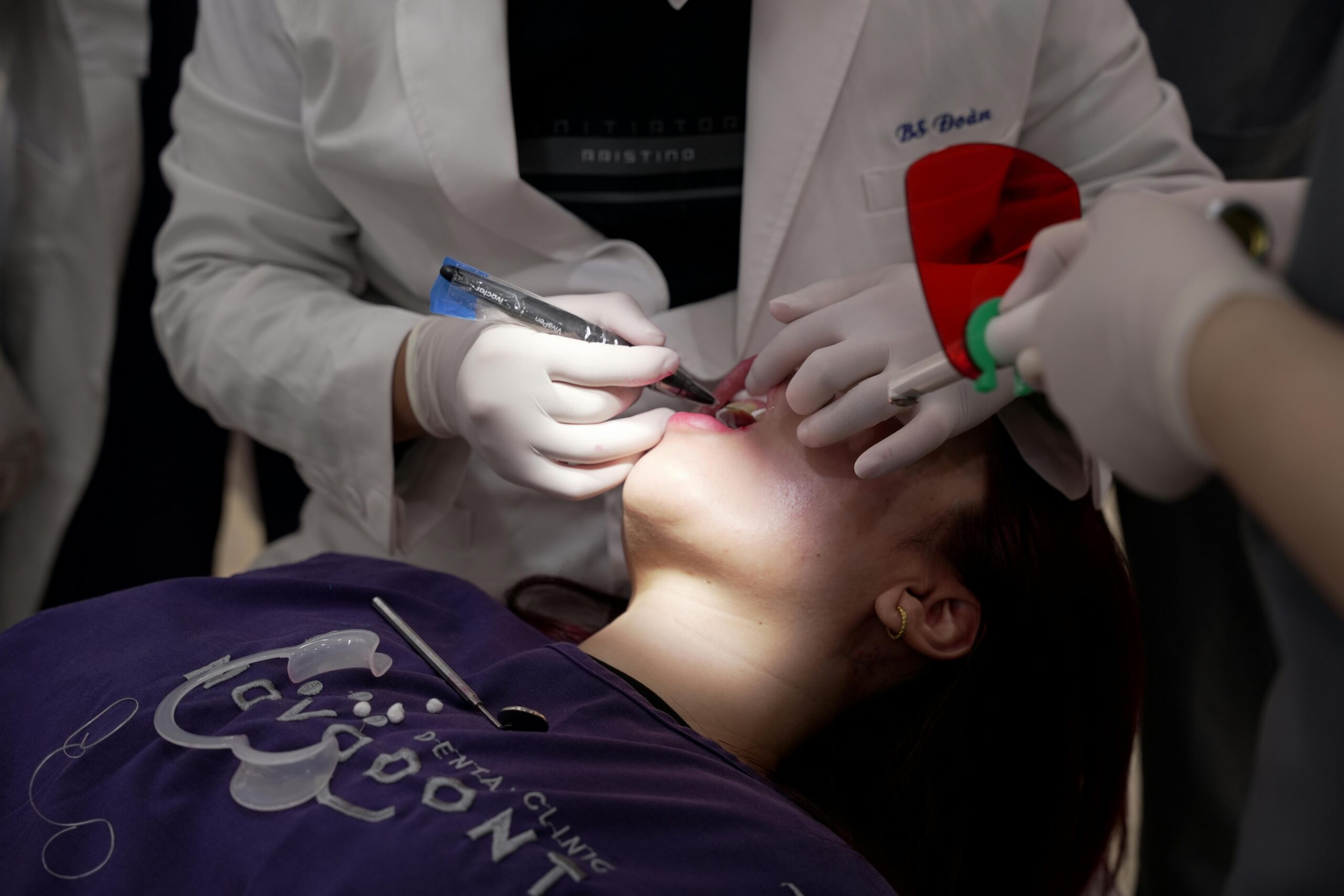
Real‑world frame: a child needs 3D imaging for an impacted canine. In that case, we select a child‑specific protocol, limit the field to the small region of interest, and shorten scan time. Good positioning and coaching help the child stay still, so one precise scan is enough. Clear planning prevents retakes and keeps exposure appropriate for age and anatomy.
The practical safeguards below guide everyday decisions and technique. Each reduces unnecessary exposure without losing the details the dentist needs.
- Justify the scan only when 3D data will change diagnosis or treatment.
- Use the smallest field of view that answers the question.
- Select low‑dose or pediatric protocols when they still meet image needs.
- Position and stabilize carefully, and coach stillness to avoid motion blur.
- Remove glasses, earrings, and loose appliances to reduce artifacts.
- Use preview positioning tools to confirm alignment before the exposure.
- Apply shielding only when it will not obscure the target; protect eyes by adjusting head tilt and beam path.
Clinical judgment matters at each step. We consider sensitivity of nearby tissues like the thyroid and eye lens, prior imaging, and whether 2D views could suffice. If the patient is pregnant, nonurgent scans are deferred; if essential, we narrow the field and optimize settings. These habits anchor cbct radiation dose safety in everyday care.
Quality systems support this approach. Teams follow checklists, verify protocols, and keep equipment calibrated so dose and image quality remain consistent over time. Images are reviewed immediately, and if a concern arises, the team troubleshoots before considering any repeat. Next, we outline how protocol design and routine quality checks are applied step by step. The safest image is the one that answers the question the first time.
Ensuring Safety in Imaging
Safety starts before the scan is ordered. We image only when it can improve diagnosis or treatment, and we match the scan to the smallest area and settings that meet the goal. During the scan, built-in controls, trained staff, and clear protocols work together to keep exposure appropriate while preserving diagnostic quality.
Before any CBCT, we confirm the clinical question and check prior images so we do not repeat information. If a patient could be pregnant, we defer nonurgent imaging or adapt the plan after discussion. We verify that the field of view and protocol fit the task and the patient’s size. A brief pre-scan pause confirms positioning, removes objects that could cause artifacts, and avoids the need for a second scan. This stepwise approach supports cbct radiation dose safety in daily care.
Safety also depends on consistent quality control. The unit is calibrated at routine intervals, and test images are taken with phantoms to confirm that detail and noise meet standards. Protocols are reviewed and updated, then locked to prevent accidental changes. Dose information is recorded, which helps compare exams over time and refine settings. Periodic case reviews ask whether each scan was justified and whether a lower dose option could have answered the question.
Patients can expect clear instructions, gentle positioning supports, and a short, quiet scan. We explain what we will see and why 3D detail matters for the decision at hand. Afterward, we confirm image quality on the spot so there is no surprise return for a retake. If you are planning a visit, you can check our current hours anytime. The simple goal is consistent, useful images with the lowest reasonable exposure.
Regulatory Standards for CBCT
Regulatory standards guide when CBCT is used, how it is performed, and how safety is verified. They focus on using 3D imaging only when it is needed, keeping exposure low, training operators, and checking equipment regularly. Rules and guidance come from national and state agencies, plus professional bodies. Together, they aim for clear images with responsible exposure.
Before a scan for a cracked tooth, the team confirms prior images. A core requirement is justification, meaning the scan should answer a specific clinical question that 2D images cannot. Optimization follows, using the smallest field of view and the lowest settings that still show the needed detail. Many regions also use diagnostic reference levels, which are typical dose benchmarks, often based on dose‑area product, to flag unusually high techniques and prompt protocol review [1].
Standards also require practical steps inside the clinic. Written protocols match the task and the patient’s size, with pediatric options when appropriate. Staff maintain training in radiation protection, and pregnancy screening is completed before nonurgent imaging. Quality assurance includes regular calibration and phantom test images, documentation of displayed dose indices, and periodic audits to keep techniques consistent over time. Professional guidance emphasizes justification, optimization of field of view and exposure, pediatric protocols, staff training, and ongoing quality assurance with records that support continuous improvement [2].
For patients, this means you should hear why a CBCT is needed, what area will be scanned, and how the settings fit your case. Your images and the displayed dose information are recorded, which helps the team refine protocols and avoid repeat scans. These safeguards support cbct radiation dose safety while preserving the detail needed for accurate care. The simple idea is clear: justified scans, optimized settings, and routine checks protect patients and guide better decisions.
ALARA Principle in Practice
ALARA means using the lowest radiation that still gives images good enough to answer the clinical question. In practice, we order CBCT only when 3D detail will change diagnosis or treatment, then tailor the scan to the smallest area and simplest settings that work. Example: a small 3D scan maps a nerve before placing one implant. The aim is clear images, minimal exposure, and no repeat scans.
Putting ALARA to work starts with planning. We confirm the question, check any prior images, and choose the smallest field of view that covers only the region of interest. Positioning is precise, with bite blocks and head supports to limit motion, and a preview ensures alignment before exposure. Settings are personalized to the task and the patient’s size, using pediatric or low‑dose protocols when they still meet image needs.
Optimization balances detail with dose. If fine bony anatomy is needed, we may use a smaller voxel size within a small field rather than enlarging the scan area. When broader context matters, we keep resolution modest to avoid unnecessary output while keeping the image diagnostic. Beam angle and tilt are adjusted to spare sensitive tissues like the eye lenses when possible. These choices, along with short scan times and motion control, support cbct radiation dose safety without compromising accuracy.
Verification closes the loop. Right after exposure, the image is checked on screen so positioning or artifacts are addressed immediately, not at a later visit. Removing metal items before the scan reduces streaks and makes a retake unlikely. For full‑arch planning, a thoughtfully selected larger field may be appropriate because one well‑planned scan is safer than multiple small scans across the jaw, such as during evaluation for snap‑in implant dentures.
For patients, ALARA is a promise of purpose, precision, and restraint. You should hear why the scan is needed, what area will be imaged, and how the settings fit your case. The best image is the one that answers the question the first time.
Patient Safety Measures
Patient safety comes first, from deciding if a scan is needed to how it is performed. We confirm a clear purpose, tailor the scan to your size and the smallest area needed, and prevent repeat images with careful positioning and pre-scan checks. Sensitive tissues are considered, and techniques are adjusted to avoid unnecessary exposure while keeping images clear.
Safety starts with screening. We review your history, past images, and any chance of pregnancy, then select a focused field of view and task‑matched settings. For a nervous teen, we coach breathing and use a forehead rest. The beam path and head tilt are adjusted to limit exposure to the eyes when possible, and the thyroid is kept out of the field unless it is the target. Dose indicators displayed by the unit are recorded, which helps refine protocols over time and supports cbct radiation dose safety in daily practice.
Motion control prevents retakes. Stable bite blocks, chin rests, and gentle head supports reduce blur, and we remove glasses and jewelry to limit streaks. Clear, simple instructions are given before the scan, and the team watches alignment on a preview screen before exposure. When anxiety makes stillness difficult, minimal, dentist‑guided options such as calm oral sedation for imaging comfort can help some patients hold still safely, reducing the chance of a second scan.
After the scan, images are checked immediately so issues are fixed on the spot, not at a later visit. Equipment is maintained and calibrated on schedule, and protocols are reviewed so settings remain consistent and appropriate. You can expect clear explanations about why the scan is needed, what area will be imaged, and how we will keep exposure low for your specific case. The right image, done right the first time, is the safest path.
Advances in Imaging Technology
Modern CBCT combines better sensors, faster mechanics, and smarter software to give clearer images with less radiation. New systems shape the beam to the target, shorten exposure time, and refine how data are rebuilt into 3D views. Together, these improvements help dentists see fine details while protecting nearby tissues. The goal is precise information with minimal exposure.
Example: a short, focused scan maps a canine near the sinus. High‑sensitivity flat‑panel detectors and low‑noise electronics capture more useful signal, so fewer X‑rays are needed to see small structures. Faster rotation and brief, pulsed exposures cut the time you must hold still, which reduces motion blur and the chance of a retake. Intelligent gantry paths and careful collimation limit the beam away from sensitive areas, such as the eye lenses, whenever anatomy and the clinical question allow.
Software has advanced as well. Model‑based and iterative reconstruction can reduce image noise at lower settings, improving clarity without pushing output higher. Metal streaks are handled more gracefully by improved artifact‑reduction tools that preserve edges and bone detail. Motion‑correction algorithms analyze subtle shifts and stabilize the image, which helps when perfect stillness is hard. Preview “scout” views and automated positioning checks confirm alignment before exposure, so the field of view stays tight and purposeful.
These design gains are paired with smarter protocols. Low‑dose and pediatric presets keep output appropriate for patient size, and voxel choices are matched to the task rather than defaulting to the highest resolution. Auto‑exposure control can adapt kV and mA to the region being scanned, maintaining diagnostic quality across thicker and thinner areas. For patients, that means fewer repeat scans, sharper images of the area that matters, and consistent cbct radiation dose safety built into everyday care. Next, we connect these tools with step‑by‑step protocols that keep dose low while preserving the details needed for accurate diagnosis and treatment.
Interpreting CBCT Results Safely
Safe interpretation means we review the entire 3D scan in a structured way, not just the area of concern. We confirm the image answers the clinical question, correlate it with your symptoms and exam, and explain what the findings mean for treatment. If something unexpected appears, it is documented and addressed or referred appropriately.
We start by verifying patient details, orientation, and that the field of view matches the request. Window and level settings are adjusted so bone, teeth, and soft tissues are seen clearly. The scan is then read in three planes, scrolling through every slice, with measurements taken near nerves, sinuses, or other sensitive areas. Prior images are compared to avoid misinterpretation, and key views are annotated for the record. Example: after a scan for an implant site, we trace the nerve canal.
Incidental findings are common, so we look beyond the main problem to check sinuses, jaw joints, airway space, and visible neck areas. Findings are categorized as expected anatomy, variations, or possible pathology, then triaged for monitoring, treatment, or specialist review. When needed, we consult an oral and maxillofacial radiologist for a formal report. If metal streaks or motion limit clarity, we first adjust viewing settings or supplement with a simple 2D image rather than repeating 3D, which supports cbct radiation dose safety by avoiding unnecessary rescans. Results may shape next steps, such as assessing cracks or bone support before planning complex crowns or bridges.
Patients should expect a clear summary: what was scanned, what was found, what matters now, and what does not. We discuss risks and options, including when no treatment is needed and when follow‑up is wise. All interpretations, measurements, and recommendations are recorded so your care team stays aligned. Next, we outline how these findings are communicated across your providers and how follow‑up imaging is timed only when it benefits your care. The safest interpretation is thorough, documented, and shared.
Frequently Asked Questions
Here are quick answers to common questions people have about CBCT and Radiation: Safety by Design in Glendale, AZ.
- What is CBCT and why is it used in dentistry?
Cone Beam Computed Tomography (CBCT) is an advanced X-ray technology that creates detailed 3D images of the teeth, jaws, and surrounding structures. Unlike standard 2D X-rays, CBCT offers more precise visualization of complex dental structures, making it valuable for procedures like implant planning, root canal treatments, and assessing impacted teeth. Dentists use CBCT to gather important information that isn’t visible with regular X-rays, ensuring comprehensive diagnosis and treatment planning.
- How does CBCT help minimize radiation exposure?
CBCT minimizes radiation exposure by tailoring the scan settings to the specific clinical need. The use of smaller fields of view limits the exposure to the area of interest. Parameters like voxel size, scan time, and exposure settings are chosen carefully to keep the dose as low as reasonably achievable. Modern CBCT units also use pulsed X-rays and refined beam collimation to reduce unnecessary exposure while maintaining image quality.
- Is CBCT safe for children?
CBCT can be safely used in children by employing child-specific protocols. This includes minimizing the field of view to the area of interest and using lower dose settings. Shorter scan times and careful positioning help ensure safety. These adjustments provide the necessary diagnostic information with minimal exposure, making CBCT a safe option when required for children’s dental care.
- How does CBCT radiation dose compare to other forms of imaging?
CBCT radiation doses vary based on scan size and settings. A small, focused CBCT scan can be similar to or slightly higher than a panoramic X-ray. Larger scans, which see broader areas like both jaws or sinuses, can have higher radiation than several small 2D images but remain significantly lower than a medical CT of the head. Matching the scan size to the clinical question is essential for balancing detail and exposure.
- What safety protocols are in place for CBCT imaging?
CBCT imaging safety is ensured through several protocols: scanning only when it adds valuable information, limiting the scan’s field of view, using low-dose settings, and positioning aids to minimize retakes. Pre-scan checks for patient positioning and removing metal items that could cause artifacts are also conducted. Equipment is regularly calibrated, and dose readings are recorded to continually refine protocols.
- How does everyday radiation exposure compare to a CBCT scan?
Daily activities expose us to small amounts of natural radiation from the ground, air, and space. A CBCT scan adds to this background, but its contribution varies with scan size and settings. A focused dental CBCT scan’s radiation dose is small compared to the cumulative background dose we all receive from daily life, such as during air travel or living at high altitudes.
References
- [1] Diagnostic reference levels for dental cone-beam computed tomography: current state and way forward. (2025) — PubMed:40795533 / DOI: 10.1016/j.ejmp.2025.105072
- [2] Optimizing radiation safety in dentistry: Clinical recommendations and regulatory considerations. (2024) — PubMed:38300176 / DOI: 10.1016/j.adaj.2023.12.002