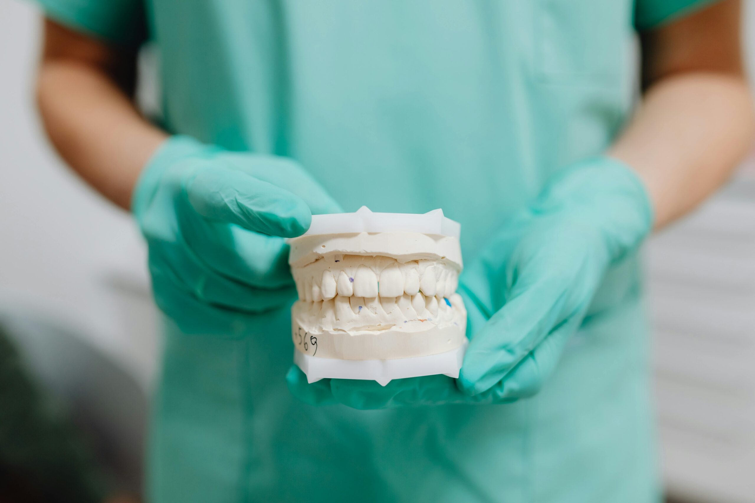
Case Overview
This case involves vertical guided bone regeneration at tooth #3 followed by implant placement. After years without an upper right first molar, chewing became uneven. The ridge had lost vertical height, so we rebuilt bone first, then placed a single implant once the site healed. In short, this vertical bone regeneration case shows a staged plan to restore function.
At the first visit, clinical exam and CBCT showed a thin, vertically deficient ridge near the maxillary sinus. Because the deficiency was vertical, we prioritized space creation and stability. The site was prepared with decortication to stimulate blood supply, particulate grafting to fill the defect, and a rigid barrier to maintain space. Tension-free primary closure protected the graft during early healing. Comfort was managed with local anesthesia, and, when appropriate, comfortable oral sedation options were discussed to reduce anxiety. After sufficient healing, we re-entered to confirm bone gain and plan implant positioning.
Implant placement focused on achieving primary stability within the regenerated bone and avoiding the sinus. Depending on soft tissue thickness, a submerged or healing abutment approach was chosen to protect the site. Provisionalization was delayed until the implant was stable, then used to shape the emergence profile. Key objectives were:
- Rebuild vertical height to support an implant of appropriate length.
- Maintain graft space with a stable barrier and minimal micromotion.
- Achieve tension-free soft tissue closure for predictable healing.
- Protect the sinus and vital structures during all steps.
- Develop healthy keratinized tissue for long-term maintenance.
For the patient, this approach aimed to restore chewing on the right side with a single-tooth implant and a custom crown. Good home care and regular follow-ups were essential during healing and after loading. When the implant integrated, a final crown completed the restoration, similar in care to other dental crowns and bridges. For practical planning and visit timing, please check our current hours. Careful planning makes vertical rebuilding possible.
Initial Presentation
At dinner, chewing on the right side felt weak. The patient arrived missing the upper right first molar (#3) for several years, with food trapping and uneven bite forces. Clinical exam and a 3D scan showed not enough bone height where the tooth was lost. From the start, this was a vertical bone regeneration case, so rebuilding height had to come before an implant.
We began with a thorough review of health history, including diabetes, smoking, sinus concerns, and any medicines that affect bone. Gum health and home care were checked to rule out active infection. Next, we mapped the ridge in three dimensions to see width, height, and the exact location of the sinus floor. The scan confirmed a vertical defect pattern rather than a simple width problem, which changes how we create and protect space for new bone.
Bite analysis followed. We looked for heavy contacts, clenching, or grinding that could overload a graft or a future implant. Soft tissue quality was also noted, since stable keratinized gum helps protect the area during healing. Adjacent teeth were tested for vitality and periodontal support to ensure they offered good neighbors and landmarks. With these findings, we discussed options, such as sinus elevation, short implants, or staged vertical rebuilding, and chose the path that best fit the defect and the patient’s goals.
Pre-surgical steps focused on risk reduction. Any gum inflammation was addressed first, and we reviewed gentle cleaning methods around the site. If grinding was present, we planned a night guard after surgery to reduce forces during healing. We also set clear expectations about time, since vertical rebuilding needs protection and patience to succeed. This careful plan allowed us to move forward confidently, knowing the bone and soft tissue conditions were defined and the goals were realistic.
With the initial assessment complete, we were ready to design the surgical approach to rebuild height and prepare for a stable implant. Thoughtful planning at the first visit improves predictability later.
Diagnostic Workup
Diagnostic workup confirms that vertical augmentation at #3 is safe and predictable. We combine medical history, a focused sinus assessment, detailed 3D imaging, and prosthetic planning to decide if height can be rebuilt and an implant placed reliably. This step sets the rules for the surgery and the timing that follows.
At the consultation, the patient asked, “Do I have enough bone?” We begin by identifying factors that affect bone healing, such as diabetes control, smoking, and medicines that influence bone metabolism. Sinus history, allergies, and prior infections are reviewed because the site borders the maxillary sinus. If the medical picture raises questions, we coordinate with the physician before scheduling grafting. This health screen informs risk, antibiotic decisions, and post‑operative protection plans.
Soft tissue is examined next. We measure mucosal thickness, the width of keratinized gum, and vestibular depth to predict closure quality. Thick, stable tissue helps seal the graft, while thin or mobile tissue increases the chance of exposure. We also note frenum pulls and scar lines, since they can add tension. These findings guide flap design and whether soft tissue augmentation should be staged or combined.
Imaging focuses on precision. A CBCT maps residual bone height, ridge width, sinus floor level, and any sinus septa that could complicate access. We locate the palatal wall, evaluate the contour of the buccal plate, and check the distance to adjacent roots to protect them. A prosthetic wax‑up or digital plan defines the ideal crown position, then we back‑plan the implant and measure how much vertical gain is needed. Because this is a vertical bone regeneration case, we pay special attention to space creation and stability, since those determine whether new bone can form where height is missing.
Finally, bite forces are reviewed. Signs of clenching or grinding shape the plan for splints and pressure control during healing. With risks mapped and goals set, we outline the surgical steps, healing intervals, and checkpoints so the patient knows what to expect. A careful workup makes the surgical plan more predictable.
Treatment Plan
The plan used two stages: rebuild missing bone vertically at #3, then place a single implant in the new bone. During meals, chewing stayed mostly on the left side. Because this is a vertical bone regeneration case, we focused first on predictable height gain, then on careful implant placement and protected healing. Each step had clear checkpoints before moving forward.
Stage one rebuilt height. A precise flap allowed good access and blood supply. Small openings in the outer bone encouraged bleeding from the marrow, which helps the graft mature. We created a stable, protected space with particulate graft and a rigid barrier, often supported by tenting screws, so the new bone had room to form without collapse. Closure was tension free, with sutures placed to keep the site sealed. After surgery, the patient avoided pressure on the area, kept the mouth clean with gentle rinses as directed, and returned for early checks to confirm the tissue stayed closed. Healing time for vertical gain was planned in months, with no forces on the ridge while bone formed.
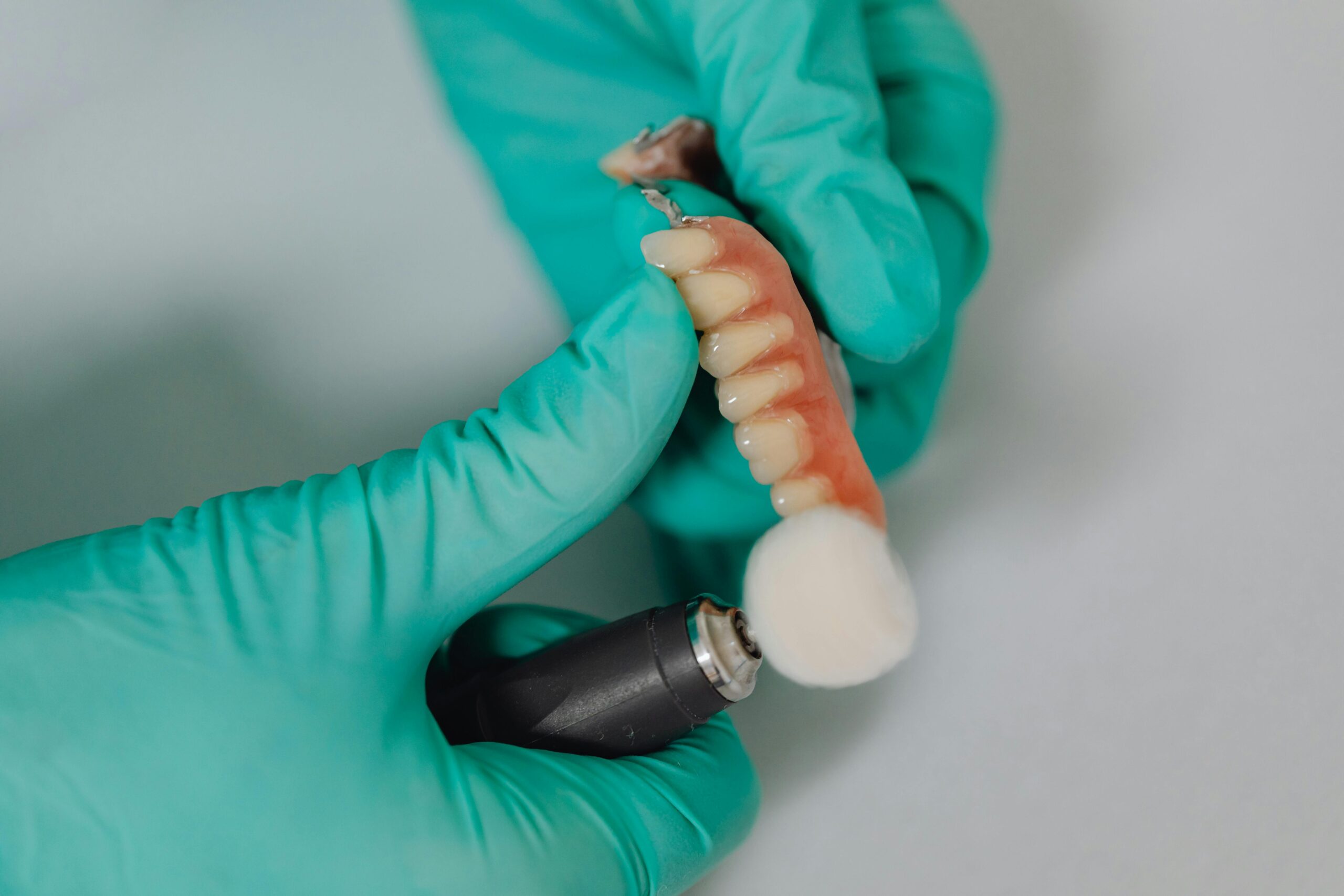
Re‑entry came after the site felt stable and imaging showed height gain. We measured the new ridge, then positioned the implant to match the planned crown and to respect the sinus. Primary stability guided the choice of a submerged cover or a small healing cap. If the tissue was thin, soft tissue shaping was planned at a later visit. The implant healed without biting forces, then a provisional crown helped shape the gum contour before the final crown. Follow‑ups confirmed comfort, cleanability, and balanced bite contacts before full use.
For the patient, this plan meant fewer surprises and steady progress. Home care stayed simple, with soft brushing and no pressure on the area. If grinding was noted, a night guard protected the work once healing allowed. With the plan set, the next step is the surgical execution that makes each checkpoint possible. Careful staging improves predictability.
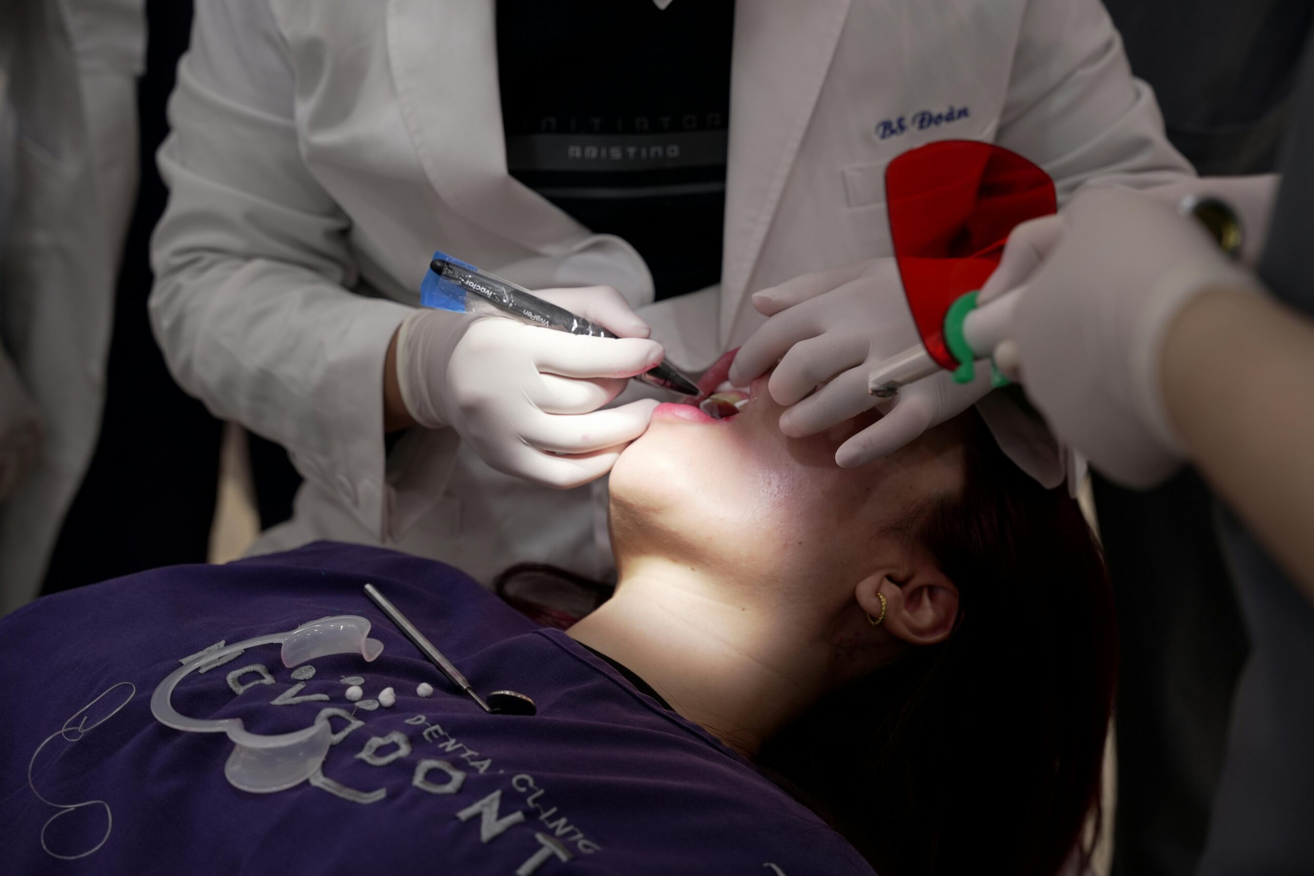
Procedure Highlights
Here is how we rebuilt vertical bone at #3 and placed a stable implant. The procedure focused on creating and protecting space for new bone, then placing the implant into that regenerated site. Each step aimed for stability, gentle handling of tissue, and a clean, sealed environment.
Picture biting into toast and feeling the right side collapse. Access began with a precise flap and careful preservation of blood supply. Small perforations in the outer cortex encouraged bleeding, which supports graft maturation. We shaped a protected, rigid space using particulate graft beneath a reinforced barrier, with fixation to prevent micromotion. Periosteal releasing incisions allowed tension‑free closure, and layered sutures sealed the site so the barrier stayed covered during early healing.
Healing occurred over months, with no pressure on the area and regular checks for quiet, intact tissue. At re‑entry, we confirmed vertical height and bone quality, then positioned the implant to match the planned crown and to respect the sinus. Primary stability guided whether the implant healed submerged or with a small cap. If soft tissue was thin, we added grafting or staged contouring to build a durable band of keratinized gum. This vertical bone regeneration case used delayed provisionalization to shape the emergence profile once the implant was stable.
For comfort, local anesthesia was standard, and longer procedures could include deep sedation options when appropriate and safe. After implant integration, a provisional verified cleanability and bite before the final crown. Follow‑ups focused on home care, gentle hygiene at the margins, and balanced contacts to protect the new work. These highlights show a simple idea carried out carefully: create space, keep it stable, and add the implant when the site is ready.
Vertical Bone Regeneration Techniques
Vertical bone regeneration techniques are methods used to rebuild lost bone height so an implant can be placed safely. Common options include guided bone regeneration with a rigid barrier, onlay or interpositional block grafts, and distraction osteogenesis. In the posterior maxilla, a sinus floor elevation may be combined when needed. The choice depends on defect size, soft tissue quality, nearby anatomy, and healing risks.
Picture a molar missing for years and the ridge too short for an implant. Guided bone regeneration creates a stable, protected space for bone to grow. Small channels in the outer bone bring blood, particulate graft fills the gap, and a supported membrane or mesh holds the shape while the body replaces grafted areas with new bone. Tension-free closure keeps the site covered, and healing is measured in months, not weeks. Systematic reviews that compare vertical augmentation methods summarize outcomes and complications, and they consistently highlight space maintenance and soft tissue management as keys to success [1].
When defects are larger or three-dimensional, block grafts or a shell approach can build vertical walls first, then fill the space with particulate graft. Thin cortical plates can be used to form a “box,” which supports the graft and preserves volume while it matures. An allogeneic shell technique has been reviewed and reported as clinically effective in selected ridge augmentation cases when protocols are followed [2]. Distraction osteogenesis, which slowly moves a bone segment upward to generate new bone beneath it, is another option for specific indications, though it requires strict patient cooperation.
For our vertical bone regeneration case at #3, we selected the least invasive method that could reliably create height and allow a well‑positioned implant. Your plan may differ based on health, tissue thickness, and how much height is needed. No matter the technique, careful planning, gentle surgery, and patient protection during healing determine the final result. The goal is simple, safe bone gain that supports a long‑lasting implant.
Impact of GBR on Implant Success
Guided bone regeneration (GBR) can improve implant success by rebuilding missing bone so the implant can be placed in the ideal position with solid support. When height or width is lacking, GBR adds volume around the future implant, which helps with stability and long‑term maintenance. Outcomes still depend on precise technique, healthy soft tissue, and careful follow‑up.
At breakfast, the right side still avoided pressure. GBR affects success in two main ways. First, it allows prosthetically driven implant placement, so forces travel down the long axis of the implant rather than into thin outer bone. Second, it creates adequate bone thickness around the implant, which supports healthy bone remodeling and easier hygiene. When the implant is positioned correctly and surrounded by stable bone, the restoration tends to function more quietly.
Vertical defects near the maxillary sinus add complexity. In a vertical bone regeneration case, rigid space maintenance and tension‑free closure protect the graft during early healing, which lowers the chance of exposure and graft loss. Staging the implant until the regenerate matures helps achieve primary stability without over‑preparing the site. Delayed loading lets bone adapt before the implant takes chewing forces, reducing early mechanical stress.
Patient factors also matter. Good gum health, no active infection, and control of habits like smoking or night grinding improve predictability. Cleanability is planned from the start, using a crown shape and emergence profile that allow daily care. Periodic checks confirm that the tissue seal stays intact and the bite is balanced, because heavy contacts can inflame the site even when bone looks perfect on a scan. Small adjustments at these visits protect the work you cannot see.
For patients, GBR can change a site that could not hold an implant into one that supports a comfortable, durable tooth replacement. When the foundation is built well, the implant has a better chance to last. Thoughtful planning and protection drive success.
Post-Procedure Care Guidelines
After vertical GBR and implant work at #3, protect the site, keep it clean, and avoid pressure while it heals. For the first 72 hours, expect mild swelling and a dull ache that improve with simple care. Follow these steps closely so the graft and implant stay stable and comfortable.
The evening after surgery, you feel mild swelling and a dull ache. Bite gently on the gauze for the first hour to control oozing, then change it as needed until bleeding slows. Use cold packs on the cheek, 15 minutes on and 15 minutes off, for the first day. Sleep with your head elevated for two nights. Do not spit, rinse hard, use straws, or smoke, since suction and smoke can disturb the clot and the graft. Take pain medicine only as directed, and finish any antibiotics if they were prescribed.
Oral hygiene starts 24 hours after surgery with gentle saltwater rinses or the prescribed rinse, twice daily. Brush the other teeth as usual, but keep the brush off the surgical area until we say it is safe. When brushing near the site is allowed, use a very soft touch and avoid the stitches. Eat soft, cool foods on the opposite side for several days. Skip hard or seedy foods that can press into the area. Do not wear or bite against a removable partial or retainer over the graft unless we have relieved and lined it to remove pressure.
Because #3 is close to the sinus, follow sinus precautions for at least two weeks. Do not blow your nose. If you must sneeze, keep your mouth open. Avoid heavy lifting or straining that makes facial pressure rise. Use only the decongestants or allergy medicines we recommend, and sip water often to keep the mouth moist.
Swelling usually peaks at 48 to 72 hours, then fades. Bruising can take a week to clear. Stitches are typically removed around two weeks, and we will check the site regularly after that. Call if you have worsening pain after day three, fever, foul taste, pus, persistent bleeding, or if you see a shiny barrier peeking through the gums. Careful daily habits protect the vertical bone regeneration case and support a long‑lasting implant.
As healing continues, we will guide hygiene around the healing cap and later the final crown so chewing feels natural again. Small steps each day add up to a quiet, durable result.
Outcome & Follow-Up
The outcome was a stable implant at #3 supported by newly gained vertical bone and healthy gum tissue. Chewing strength returned to the right side, and the final crown felt natural and easy to clean. The sinus remained intact, and the bite was balanced without heavy contacts.
At the six-month visit, biting toast felt even again. Radiographs showed maintained vertical height and bone around the implant neck. We confirmed implant stability before any provisional crown, then shaped the gum line so the final restoration would seal well and allow daily hygiene. In this vertical bone regeneration case, the key measures of success were quiet tissues, stable bone on imaging, and a crown positioned for cleanability.
Follow-up moved in clear steps. Early checks in the first two weeks focused on comfort, swelling, and keeping the incision closed. During graft healing, visits every four to six weeks monitored the soft tissue and ensured no pressure from appliances or chewing. Re-entry occurred only after the site felt firm and imaging showed height gain. After implant placement, we allowed undisturbed healing, then used a provisional to shape the emergence profile before the final crown. Each step required the previous one to be stable.
Longer term, we verified that the bite stayed light on the crown in excursions, since off-axis contacts can stress new bone. For patients who grind, a night guard was advised once healing permitted, to limit peak forces. Home care included gentle brushing at the margins and floss or interdental aids designed for implant contours. Baseline radiographs were taken at restoration, then again around 6 to 12 months to confirm stable bone levels. Routine maintenance visits kept the tissue seal healthy and allowed small adjustments before problems developed.
For the patient, this plan meant steady progress and a tooth replacement that felt like part of the mouth, not a foreign object. Clear checkpoints and simple daily habits protected the result and supported long-term comfort. Small, consistent steps keep the implant quiet.
Lessons Learned from the Case
Three lessons stood out. Plan the implant position first, then build bone to fit that plan. Protect the graft from pressure and movement. Move in stages, and let biology set the pace.
Start with diagnosis, not tools. A careful CBCT and prosthetic plan showed the defect was vertical, near the sinus, so space creation and rigid protection mattered more than speed. We learned to target the smallest augmentation that achieves the crown-driven position, rather than trying to rebuild extra height “just in case.” Safety margins around the sinus and adjacent roots kept the site calm and reduced surprises. In this vertical bone regeneration case, success came from matching the regenerate to the final tooth, not the other way around.
Soft tissue decided predictability. Thick, healthy gum sealed the site and kept barriers covered; thin tissue risked exposure. Flap design, gentle handling, and true tension-free closure were more important than any single material. Patient steps mattered too. No biting on the area, no pressure from appliances, and simple, gentle hygiene kept the clot and graft quiet. A small real-world moment: avoiding chewy bread on the surgical side saved the early seal.
Timing and checkpoints guided every move. Re-entry was based on firm palpation and imaging, not the calendar. Implant stability set whether we submerged or used a small healing cap. We delayed the provisional until the implant felt solid, then used it to shape the gum for easy cleaning. Bite forces were kept light, with adjustments after delivery and a night guard for grinders when healing allowed. These habits kept the new height stable long term and made daily care simple for the patient.
For patients, the takeaway is clear. Careful planning, gentle surgery, and steady protection turn a thin ridge into a strong, comfortable tooth site. Plan slow, protect space, and let biology work.
Frequently Asked Questions
Here are quick answers to common questions people have about Case Report: Vertical GBR and Implant at #3 in Glendale, AZ.
- What is vertical bone regeneration?
Vertical bone regeneration is a dental technique used to rebuild lost bone height in the jaw. This process is essential when the bone height has decreased, making it impossible to place dental implants properly. The techniques often involve guided bone regeneration with a membrane to create and maintain space, allowing new bone to grow over time. This procedure can take several months, but it provides a stable foundation for future dental implants, helping to restore both function and aesthetics.
- How is guided bone regeneration (GBR) performed?
Guided bone regeneration (GBR) is performed by creating a stable and protected space in the jaw, often using a barrier membrane and bone graft material. The process involves making small openings in the bone to encourage blood supply, filling the area with graft material, and covering it with a barrier to maintain the desired shape. The goal is to allow new bone to form in the space while preventing soft tissue from collapsing into it. This ensures the necessary support for future implant placement.
- What makes vertical bone regeneration important before dental implants?
Vertical bone regeneration is crucial before dental implants because it rebuilds the lost bone height required to securely anchor the implants. Without sufficient bone, implants may not integrate correctly, leading to potential failure. Rebuilding bone vertically provides the necessary support and ensures that implants can be placed in the optimal position for function and aesthetics. It helps in distributing bite forces evenly and prevents complications associated with insufficient bone height.
- What are common techniques used in vertical bone regeneration?
Common techniques for vertical bone regeneration include guided bone regeneration, using either a rigid barrier with particulate graft or block grafts to recreate the missing vertical bone. Distraction osteogenesis is another technique, where bone segments are gradually moved apart to allow new bone to form in the gap. The choice of technique often depends on the specific anatomy and health status of the patient, as well as the size and shape of the bone defect.
- How long does it take to heal after vertical bone regeneration?
Healing after vertical bone regeneration typically takes several months, as the grafted material needs time to integrate and mature into new bone. During this period, it is important to avoid putting pressure on the area and to maintain good oral hygiene to prevent infection. Regular follow-ups with your dentist will help monitor the progress and ensure the graft is healing properly before any implants are placed. Patience and careful post-procedure care are key to successful healing.
- What factors influence the success of vertical bone regeneration?
Several factors influence the success of vertical bone regeneration, including the patient’s overall health, particularly control of systemic conditions like diabetes and habits like smoking. The quality of soft tissue, ensuring tension-free closure during surgery, and meticulous post-operative care are also crucial. Proper technique during the surgery and adherence to follow-up appointments ensure the newly regenerated bone provides a stable, durable site for future implants.
- Can vertical bone regeneration help with sinus issues?
Yes, vertical bone regeneration can help with sinus issues, especially when the missing tooth is in the upper jaw near the sinus cavity. Sometimes, the procedure includes sinus floor elevation to create enough vertical space and facilitate implant placement. By carefully planning and executing the procedure, potential complications with the sinus, such as perforations, are minimized, allowing for a smoother and more predictable outcome.
References
- [1] Evaluating Techniques for Vertical Ridge Augmentation via Comparative Study of Clinical Outcomes: A Systematic Review. (2025) — PubMed:41464541 / DOI: 10.3390/jcm14248639
- [2] Clinical Efficacy of Allogeneic Bone Plate Shell Technique for Alveolar Ridge Augmentation: A Systematic Review. (2025) — PubMed:41552114 / DOI: 10.7759/cureus.99228