Understanding CBCT Technology
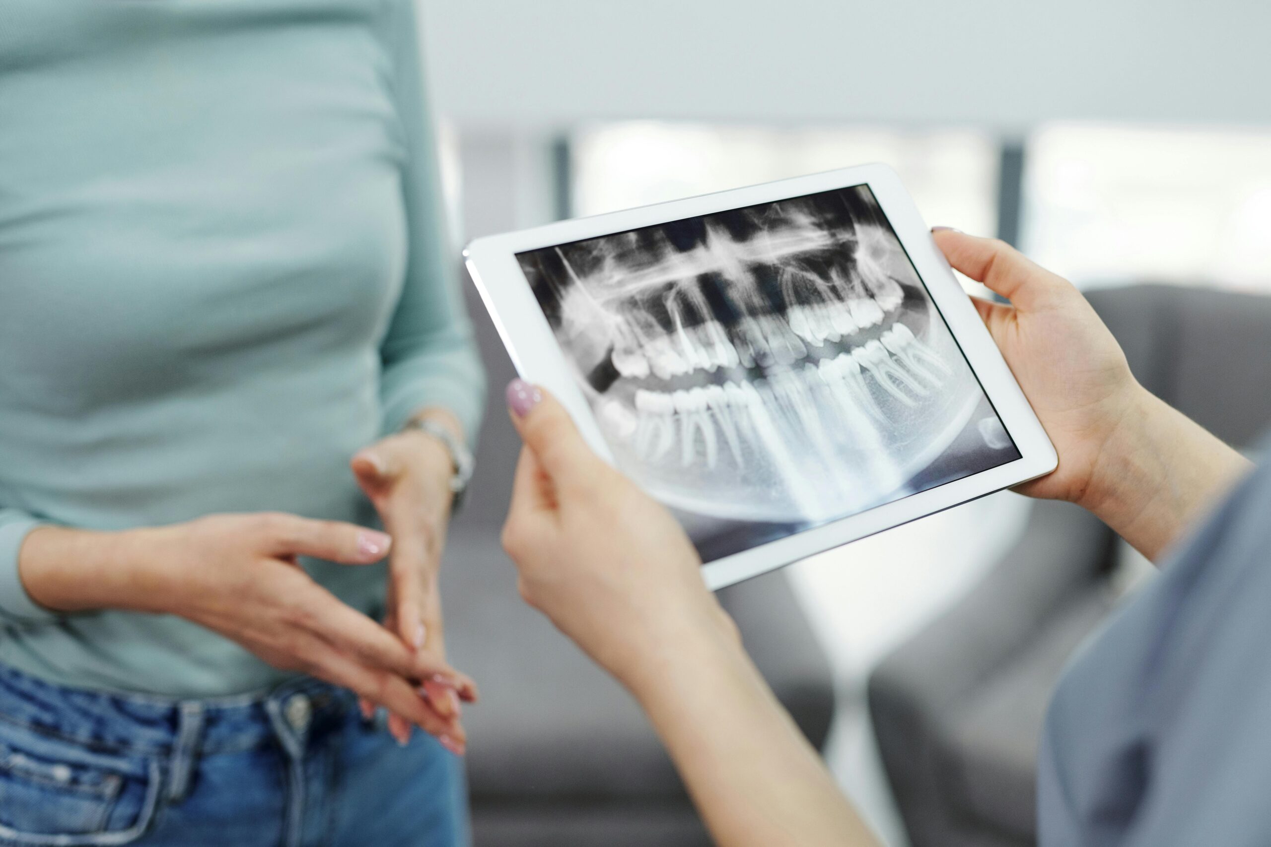
CBCT, or cone beam computed tomography, is a 3D dental X-ray that shows teeth, jaws, nerves, and sinuses in detail. The machine rotates around your head, taking many low-dose images, then a computer builds a 3D model. This view helps us see depth and exact positions that plain X-rays cannot. For scheduling, see our current hours.
Compared with 2D images, CBCT provides cross-sections and true measurements. For implant planning, it shows bone height and width, and the relationship to the maxillary sinus or nerve canals, which helps avoid surprises and guides safer placement [1]. It also supports analysis of the peri-implant phenotype to match tissue and bone conditions with the planned restoration [2]. Before an implant, we map your jaw in 3D. Many now view CBCT as cbct essential modern dentistry for precise diagnosis and planning, whether for single-tooth implants or full-arch implant dentures.
Common times we use CBCT include:
- Planning dental implants near the sinus or major nerves.
- Evaluating complex root canal anatomy or suspected hidden canals.
- Locating impacted teeth and their position to roots and nerves.
- Assessing jaw joints and bite-related bone changes.
- Reviewing sinus health and airway space during dental care.
- Checking fractures or jaw injuries after trauma.
For patients, CBCT often means fewer appointments, clearer plans, and more predictable care. It can reveal hidden canals and infection before root canal treatment, which guides gentler access and improves precision. Next, we will build on this foundation to show how these images inform step-by-step treatment choices. Clear 3D images support safe, confident care.
Importance of 3D Imaging
3D imaging matters because it shows depth and exact positions that flat X-rays cannot. With a true three-dimensional view, we see teeth, bone, nerves, and sinuses as they actually relate to each other. This reduces guesswork, supports safer decisions, and helps tailor treatment to your anatomy. Picture a hidden root curve that only appears when we can see around it.
The key advantage is accuracy. CBCT creates a volumetric model that allows precise measurements in any direction, so we can judge bone thickness, locate nerve canals, and map sinus walls before any procedure. Subtle problems, such as fine cracks or early bone changes, may be clearer in 3D. This clarity improves planning, from choosing the least invasive path to selecting the right instruments and angles. It also improves communication, since we can show you the area and explain options with confidence.
3D imaging also helps anticipate and avoid complications. For example, when evaluating a deeply positioned third molar, the scan can reveal its true distance to the nerve, guiding a gentler approach to wisdom tooth removal. In orthodontics, seeing root positions and bone contours supports aligner plans that move teeth within safe boundaries, reducing unwanted root contact or bone loss. This leads to more predictable movements and fewer mid-course corrections.
Beyond diagnosis, 3D data can be used for virtual planning and guide fabrication. Surgical or orthodontic guides translate the plan to the mouth with high fidelity, which can shorten procedures and improve fit. Exposure is managed by choosing the smallest field of view that answers the clinical question, aligning with practical dose principles. Not every case needs a scan, but when anatomy is complex or uncertainty remains, 3D imaging provides clarity that 2D views cannot. For aligner care, this often means better staging and fewer surprises; see how we plan Invisalign treatment with confidence.
In short, 3D imaging turns difficult decisions into clear, well-guided steps.
Role of CBCT in Diagnosis
CBCT helps us find the exact source of a dental problem by showing a true 3D picture of teeth and jaws. When a 2D X-ray is unclear, CBCT clarifies what is happening, where it is, and how large it is. Think of a sore tooth that still hurts after a normal X-ray. This clarity is why many consider it cbct essential modern dentistry for accurate diagnosis.
In endodontics, CBCT can separate overlapping structures and reveal the buccal or lingual location of disease. That means we can see whether a dark area comes from a cracked root, external resorption, or a lesion next to a vital tooth. It also shows the true size of an infection and its relationship to nearby bone. With that information, treatment can target the exact cause, not just the symptoms.
CBCT is also useful for surgical and periodontal diagnosis. It maps impacted teeth in three dimensions, so their position to nerves and adjacent roots is precise. In the sinus, CBCT can show membrane thickening and bony walls, which helps explain nasal-sinus contributions to dental symptoms and informs implant plans [3]. For the jaw joints, 3D imaging can display condylar shape and cortical changes that are not visible on standard films, supporting a more complete temporomandibular joint assessment [4]. When planning implant-retained dentures, 3D evaluation of bone height and sinus location guides safer, more predictable snap-in implant denture planning.
Practically, CBCT is ordered to answer a focused clinical question, not by routine. The scan should change the plan, such as confirming a fracture before retreatment or mapping a safe path for a tooth removal. For patients, this means fewer surprises, clearer choices, and procedures tailored to your anatomy. Clear diagnosis leads to confident care.
Identifying Missed Canals
Missed canals are hidden pathways inside a tooth that were not located or cleaned during treatment. CBCT helps identify them by showing the tooth in three dimensions, so canals are not hidden behind overlapping roots. With cross-sectional views, we can spot extra canals, sharp curves, and calcifications that 2D X-rays may miss. A patient still aches after a root canal; CBCT shows a hidden second canal.
Upper first molars often have an extra canal in the mesiobuccal root. CBCT studies map these additional canals and their orientations, which guides precise access and instrument selection for a safer approach [5]. When symptoms persist after treatment, a limited field-of-view scan can reveal a previously untreated canal or a small lesion around the root that was not visible on standard periapical films [6]. This added clarity reduces guesswork and helps us target the true cause of discomfort.
CBCT also helps when canals are calcified or the tooth has a large filling or crown. The 3D view shows where the canal shifts under restorations and where dentin is thin, so access can be more conservative. In retreatment, we evaluate existing fillings, post position, and any ledges or perforation risk before touching the tooth. By confirming problems like an unfilled canal or a missed isthmus, CBCT supports careful planning and reduces unnecessary tooth removal [7].
For patients, identifying a missed canal early can shorten retreatment, relieve lingering pain, and protect tooth structure. Not every case needs a scan, so we choose the smallest field that answers the question and keep dose as low as practical. This focused, evidence-based use is one reason CBCT is viewed as cbct essential modern dentistry in endodontic care. Next, we will show how 3D findings guide each step from access to sealing.
Enhancing Implant Safety Procedures
CBCT enhances implant safety by showing a true 3D map of bone, nerves, and sinuses before surgery. This lets us plan the exact implant position, length, and angle while avoiding vital structures. Before placing a lower molar implant, we map the nerve canal in 3D. The result is a safer plan with fewer surprises.
Safety starts with measurement. CBCT shows bone height and width, the course of the inferior alveolar nerve, and sinus walls with millimeter-level clarity. With that map, we test different implant sizes virtually, check safe distances, and choose a path that preserves healthy bone. When needed, we design a surgical guide from the scan so the plan transfers to your mouth with precision, which supports accuracy and reduces risk to nerves and adjacent teeth [8]. Many clinicians now consider CBCT cbct essential modern dentistry for implant planning near delicate anatomy.
The scan also informs the surgical approach. If bone is thin, we can adjust implant diameter or stage grafting to prevent perforations. If the sinus floor is low, we plan a lift only where needed and choose the least invasive technique. In immediate implants, CBCT shows socket shape and root positions, so we place the implant into solid bone rather than empty space. These steps reduce the chance of nerve irritation, sinus complications, and unintended cortical breaches.
Intraoperatively, a guide based on the CBCT plan helps control angulation and depth, promoting steady, gentle drilling. Postoperatively, a low-dose check image can confirm position when the situation warrants it, helping us respond early if adjustments are needed. Dose is managed by using the smallest field that answers the safety question, keeping exposure practical while preserving detail.
For patients, this planning means smaller incisions when appropriate, less time in the chair, and a lower chance of numbness or sinus issues. Clear 3D planning builds confidence for single-tooth and full-arch cases alike. In the next section, we will connect these safety steps to day-by-day implant healing and follow-up. Safer planning leads to smoother recovery.
Comprehensive Treatment Planning
Comprehensive treatment planning means building a clear, step‑by‑step roadmap for your care before we begin. CBCT adds the missing depth, so we can place teeth, bone, nerves, and sinuses into one accurate picture. With this 3D view, we align function, comfort, and appearance while reducing surprises.
We start by merging CBCT data with digital impressions and bite records. This creates a single model that lets us design the desired tooth positions first, then test how well bone and soft tissue can support that plan. When fixed restorations are part of the plan, we evaluate root shape, remaining tooth structure, and occlusion to support predictable results, such as thoughtful planning for crowns and bridges that fit your bite. This prosthetic‑first approach keeps the final goal in focus while checking safe distances to nerves and sinuses.
Sequencing is just as important as design. CBCT helps us decide which teeth can be maintained, which should be removed, and where grafting may be needed, all while preserving healthy bone. If a temporary replacement is needed during healing, we plan space and support for well‑fitting partial dentures during transitions. For patients who clench, we review joint spaces and condylar position to protect the plan from excessive bite forces. For esthetics, we study smile lines and bone levels so tooth shapes and gum contours look natural and remain cleanable.
From there, the plan becomes action. Surgical guides can translate the digital plan to your mouth, and restorative guides help control tooth shaping with minimal removal. If the scan shows thin bone or a low sinus floor, we stage care to stay conservative. For you, this means fewer mid‑course changes, shorter appointments, and clearer choices at each step. This clarity is one reason many view cbct essential modern dentistry for coordinated, multi‑step care. Next, we connect this planning to how procedures are carried out day to day so healing stays on track.
A precise plan makes treatment safer, smoother, and more predictable.
Benefits Over Traditional Imaging
CBCT offers clear 3D views that traditional 2D X-rays cannot provide. It removes overlap and distortion, so measurements are true to size and position. A molar aches, but standard X-rays look normal; CBCT can show a hidden crack or small cyst.
With 2D images, parts of the mouth stack on top of each other, and magnification can fool the eye. CBCT slices the area into thin sections in every plane, revealing the exact shape of teeth and bone. This helps pick safer paths for care and reduces guesswork. One scan often answers several questions at once, replacing multiple angled films. Targeted fields keep the view tight to the problem and avoid unnecessary areas.
These details support more conservative care. We can see remaining dentin before shaping a tooth, or the true distance between roots, which helps avoid over-cutting. Around teeth, the 3D view shows bone defects and their walls, guiding repair plans and graft choices. After trauma, CBCT reveals fine fractures and their direction, so treatment is precise rather than exploratory. For patients with a strong gag reflex or limited opening, the external scan is often more comfortable than holding sensors inside the mouth.
The 3D data also improves teamwork and communication. We can share the same model with surgeons, restorative dentists, and labs, so everyone plans from identical information. That coordination shortens appointments, reduces surprises, and supports guides that turn the plan into accurate steps. We order CBCT only when it is likely to change care, and we keep exposure practical by limiting the scan size. These advantages are why many clinicians consider it cbct essential modern dentistry for complex decisions. Clear images lead to confident choices.
Case Studies Demonstrating Effectiveness
Real cases show how CBCT changes decisions and improves outcomes. By revealing exact 3D anatomy, it often alters the plan before we touch a tooth. This clarity can prevent complications, shorten procedures, and reduce follow-up visits.
A patient needs a molar replaced near the nerve. The CBCT reveals a deep lingual undercut and the true path of the nerve canal, which a panoramic film could not show. We adjust to a shorter, narrower implant and select a controlled entry angle, then use a guide to reproduce that path. The surgery proceeds without nerve irritation and with minimal bone removal.
In another case, a tooth still aches months after a root canal. A small field-of-view scan shows a missed second canal and a tiny lesion hidden between roots. Knowing the exact canal location lets us create a conservative access, clean the missed space, and seal it well. Symptoms resolve, and the tooth is preserved without exploratory cutting.
A different situation involves an impacted canine close to front tooth roots. Standard X-rays cannot show whether it sits toward the cheek or the palate. CBCT pinpoints a palatal position and shows early wear on a nearby root, so the exposure and traction path are changed to avoid damage. Orthodontic movement is planned within safe bone, and long-term stability improves.
Finally, a patient has pressure in the upper jaw and chewing pain. The scan shows a thickened sinus membrane and a dental source in a back molar area. Treating the tooth first resolves symptoms, and the 3D map later guides a precisely placed sinus lift only where needed.
These outcomes come from better information, not guesswork. They explain why many clinicians view cbct essential modern dentistry for complex choices. Next, we focus on how scan settings and workflow keep dose practical while preserving detail.
Integrating CBCT in Daily Practice
Integrating CBCT into daily care means building clear steps around when, why, and how we scan. We ask a focused clinical question, choose the smallest field of view that answers it, then plan from the 3D map. Morning huddle: today’s plan flags one complex molar and an implant site. This keeps imaging purposeful, efficient, and tied to decisions.
Workflow starts chairside. We confirm need, review medical history, and explain the goal of the scan. Positioning is stabilized, motion is minimized, and exposure is tailored to the region of interest. Immediately after acquisition, the dentist reviews key cross-sections with the patient, documents justification and findings, and records settings used. This ALARA-focused routine keeps dose practical while making the image central to the plan.
Next, we integrate data. CBCT volumes are aligned with digital impressions and bite records so surgical, orthodontic, and restorative plans share the same model. When indicated, we design guides that transfer the plan precisely to the mouth. Standardized file naming and secure sharing help the team work from identical information. Emerging tools that map sinus-tooth relationships can further streamline review and reduce oversight by highlighting critical anatomy on CBCT images [9].
Quality and comfort round out daily integration. Staff follow positioning checklists, and equipment receives routine image-quality checks. Preoperative CBCT features have been associated with postsurgical pain and swelling after endodontic microsurgery, which helps set expectations and refine surgical plans for comfort [10]. For anxious patients, we coordinate care with patient-friendly oral sedation options when procedures are needed. This dependable workflow is one reason many view cbct essential modern dentistry for team-based, patient-centered care.
The result is faster decisions, fewer surprises, and plans that match your anatomy.
Future Trends in Dental Imaging
Future dental imaging will be smarter, faster, and more connected to your overall plan. Expect clearer 3D views with tools that help the dentist measure, mark risks, and simulate treatment before anything begins. Tomorrow’s visit may include a scan that plans and guides care in minutes. The goal is safer, more precise decisions with less guesswork.
Artificial intelligence is a major driver. New systems learn to spot patterns on CBCT, mapping canal pathways, nerves, and other anatomy to support endodontic and surgical planning [11]. Broader reviews describe growing clinical uses for AI across dentistry, including diagnosis, triage, and decision support that can streamline complex cases [12]. As these tools mature, they will flag risk zones, suggest measurements, and reduce time spent scrolling through slices.
Hardware and software are also advancing. Smaller, targeted fields of view, improved detectors, and motion correction aim to capture the area of interest clearly while keeping exposure practical. Image fusion is becoming routine, combining CBCT with intraoral scans and facial photos so the plan reflects real tooth shape, bite, and smile. With this unified model, the team can design surgical guides or orthodontic plans, then 3D print guides chairside for accuracy and speed. Navigation systems and simple augmented overlays are emerging to help the hand follow the digital plan more closely in real time.
These trends matter for patients because they reduce surprises and make plans easier to understand. A scan that highlights a thin bone area or a nearby nerve helps tailor implant placement and may shorten procedures. For orthodontics and esthetics, merging datasets supports tooth movement and shaping within safe, healthy boundaries. As this technology becomes routine, it reinforces why many consider cbct essential modern dentistry for confident, informed care. We will continue to translate these innovations into clear, step‑by‑step choices you can review before treatment begins.
Smarter imaging aims to make dentistry safer, faster, and more precise.
Frequently Asked Questions
Here are quick answers to common questions people have about CBCT in Modern Dentistry: Non-Negotiable in Glendale, AZ.
- What makes CBCT essential in modern dentistry?
CBCT is essential in modern dentistry because it provides detailed 3D images that reveal the exact positions of teeth, nerves, and bones. This technology reduces guesswork, leading to safer and more accurate diagnoses and treatments. It helps in planning complex procedures like dental implants, revealing hidden structures such as impacted teeth or extra root canals, and even in assessing jaw conditions. By allowing dentists to see what standard X-rays might miss, CBCT improves the precision and predictability of dental care.
- How does CBCT improve implant safety?
CBCT improves implant safety by providing a detailed 3D map of the patient’s anatomy. It shows vital structures like nerves and sinuses, allowing dentists to plan the exact position and angle of an implant without risking these areas. This reduces the chance of complications such as nerve injury or sinus problems. Additionally, CBCT aids in creating surgical guides that help in accurate implant placement, ensuring the procedure is performed with precision.
- Can CBCT help identify missed root canals?
Yes, CBCT can identify missed root canals by providing a 3D image of the tooth’s internal structures. This view helps in locating extra or hidden canals that may not be visible on a standard 2D X-ray. By showing sharp curves and calcifications, CBCT assists in more accurate endodontic treatment, reducing the likelihood of persistent pain or the need for retreatment. This technology is particularly useful in complex cases where overlapping structures could hide problems.
- Why is CBCT preferred over traditional 2D X-rays?
CBCT is often preferred over traditional 2D X-rays because it offers a true three-dimensional view of the dental structures. This prevents the distortion and overlap common with 2D images, allowing for more accurate diagnosis and treatment planning. It helps in detecting small fractures, hidden canals, and other intricate details, supporting more effective and conservative dental care. Additionally, CBCT scans can often solve multiple clinical questions in one go, limiting the need for multiple x-rays.
- How does CBCT facilitate comprehensive treatment planning?
CBCT facilitates comprehensive treatment planning by merging detailed 3D anatomical data with digital impressions and bite records. This integration allows dentists to assess the viability of treatment plans, check interferences between proposed restorations and existing structures, and tailor interventions to the patient’s unique anatomy. Sequencing care, such as deciding which teeth need removal or grafting, becomes more precise, reducing surprises and ensuring more predictable outcomes.
References
- [1] Radiographic assessment of the correlation between maxillary sinus dimensions and greater palatine canal pathway in CBCT images (a retrospective study). (2025) — PubMed:40958094 / DOI: 10.1186/s12903-025-06782-w
- [2] Three-dimensional digital quantitative analysis of periodontal and peri-implant phenotype-A narrative review. (2025) — PubMed:40641458 / DOI: 10.1111/prd.12639
- [3] Does Periodontal Bone Loss Play a Significant Role in Schneiderian Membrane Thickening? A Cone-Beam Computed Tomography Evaluation. (2025) — PubMed:41010920 / DOI: 10.3390/medicina61091529
- [4] The Combined Impact on Condylar Changes of Sex and Cortication on Mandibular Condyle: A Binary Logistic Regression Analysis. (2025) — PubMed:41018957 / DOI: 10.1155/ijod/5363346
- [5] Evaluation of root and root canal morphology in maxillary first molar teeth: A cone-beam computed tomography study using two classification systems in a Japanese population. (2025) — PubMed:40654430 / DOI: 10.1016/j.jds.2024.12.005
- [6] 2D versus 3D radiographic assessment of asymptomatic persistent endodontic lesions. (2025) — PubMed:40509944 / DOI: 10.1111/iej.14272
- [7] A Retrospective Study of CBCT-Based Detection of Endodontic Failures and Periapical Lesions in a Romanian Cohort. (2025) — PubMed:41010569 / DOI: 10.3390/jcm14186364
- [8] Dynamic navigation systems in dento-alveolar surgery: advancements and clinical applications. (2025) — PubMed:41091391 / DOI: 10.1007/s44445-025-00072-5
- [9] Enhanced diagnostic pipeline for maxillary sinus-maxillary molars relationships: a novel implementation of Detectron2 with faster R-CNN R50 FPN 3x on CBCT images. (2025) — PubMed:41015768 / DOI: 10.1186/s12903-025-06337-z
- [10] Factors Associated with Postsurgical Pain and Swelling Following Endodontic Microsurgery: The Role of Radiographic Characteristics. (2025) — PubMed:40361773 / DOI: 10.3390/healthcare13090995
- [11] Artificial Intelligence in the Study of Root and Canal Anatomy: A Comprehensive Review on Applications, Advantages, Challenges and Future Directions. (2025) — PubMed:40995722 / DOI: 10.14744/eej.2025.37232
- [12] An Analysis of Artificial Intelligence Studies in Dentistry: Clinical Applications and Next Steps. (2025) — PubMed:40960111 / DOI: 10.5005/jp-journals-10024-3867