Introduction to PRF and PRP
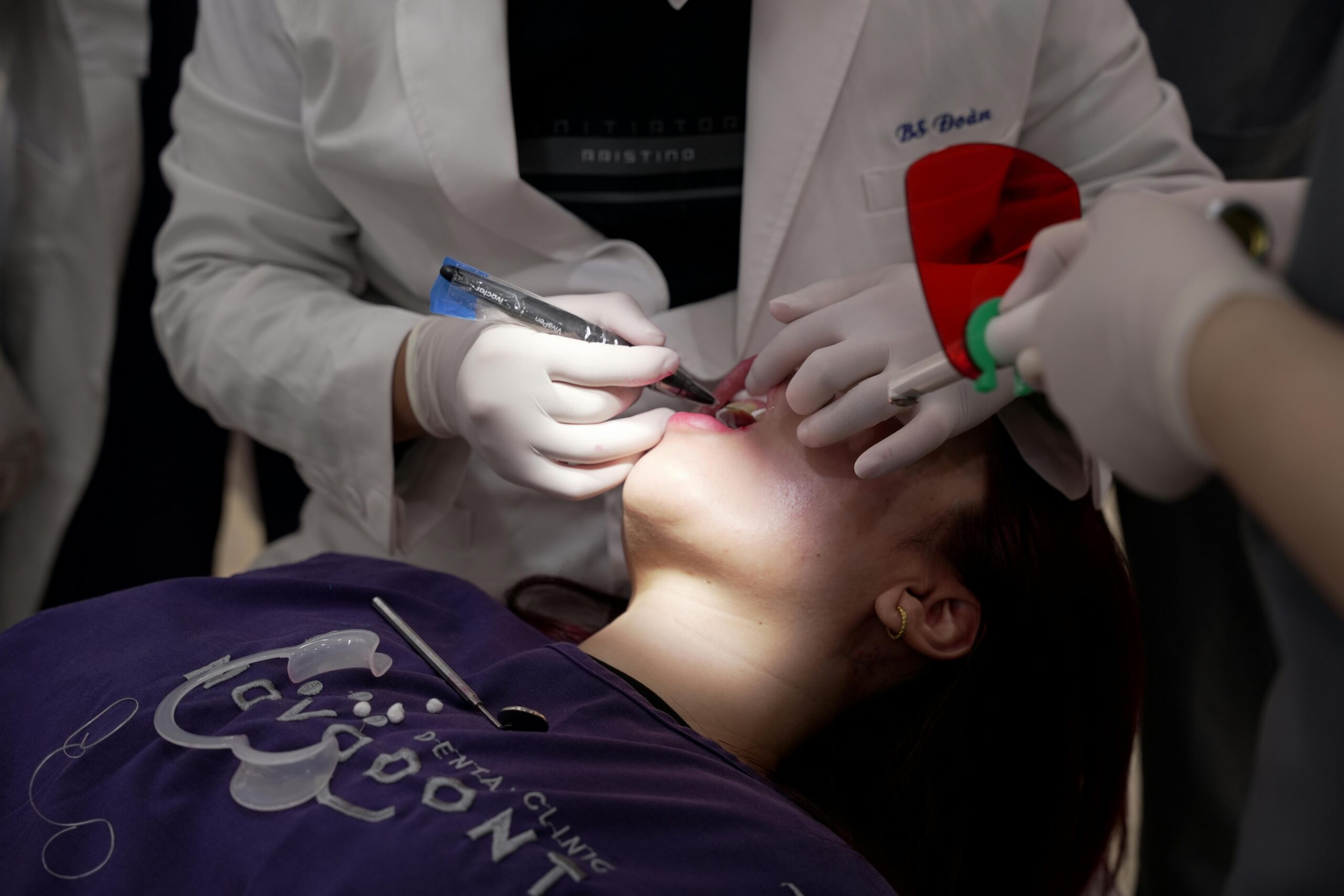
PRF and PRP are treatments made from your own blood to help healing in the mouth. Dentists use them during surgeries, grafts, and some endodontic procedures to support soft tissue and bone repair. Imagine a tooth is removed and a small blood concentrate is placed to protect the socket. If you are curious about prf prp in dentistry evidence, this overview explains what they are and why they are used.
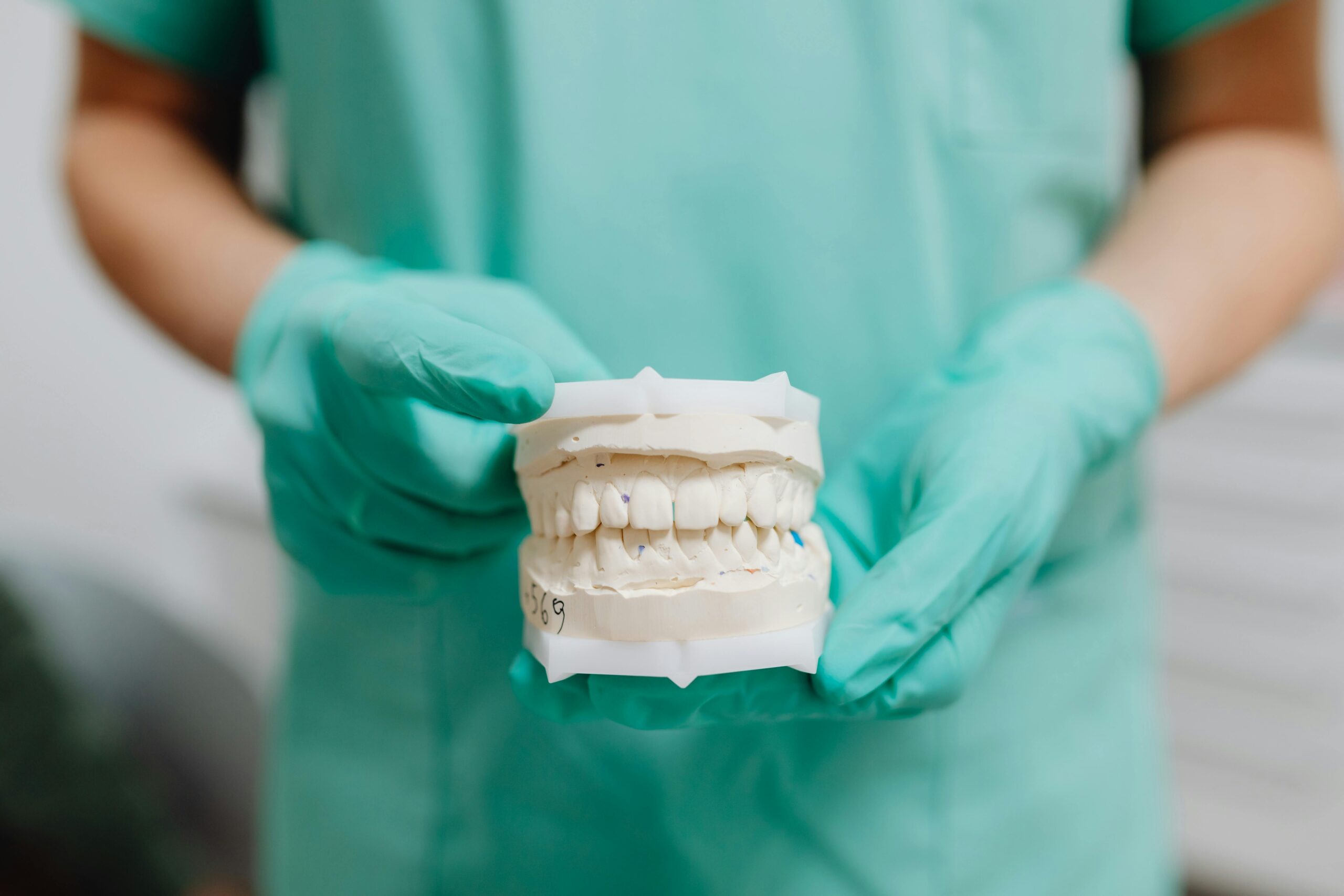
How they are made matters. A small blood draw is spun in a centrifuge to separate parts. PRP, or platelet-rich plasma, is a liquid concentrate of platelets and growth factors; it is applied or injected into the site. PRF, or platelet-rich fibrin, forms a sticky clot or membrane without added anticoagulant, which can act like a scaffold over a wound. PRF tends to release growth factors over time, while PRP gives a quicker release. These differences help guide choices in procedures such as wisdom tooth removal, where a membrane may cover the socket for comfort and protection.
What does research show so far? A recent umbrella review found potential benefits of PRF and PRP for certain dental surgeries, yet the overall certainty of evidence is limited by small, varied studies [1]. In endodontics, a scoping review mapped uses and noted promising applications alongside the need for standardized protocols and stronger trials [2]. These topics often intersect with root canal treatment and related regenerative procedures, so discussions focus on your specific diagnosis and goals.
For patients, PRF and PRP are autologous, so allergy risk is low, but results still depend on technique and case selection. In the next section, we will look at how they are prepared in the clinic and where each may fit best. They are tools, not magic; benefits depend on the procedure and technique.
How PRF and PRP Work in Dentistry
PRF and PRP work by concentrating your platelets and forming a biologic scaffold at the treatment site. During implant placement, a small PRF plug is pressed around the fixture. The platelets release signals that help stop bleeding, build new blood vessels, make collagen, and start bone repair. PRF also provides a stable matrix that cells can attach to, while PRP bathes the area with a quick burst of signals.
At the tissue level, activated platelets degranulate, releasing PDGF, TGF-beta, VEGF, and other cytokines. These signals recruit cells, spur angiogenesis, and guide early bone and soft tissue repair; the fibrin matrix acts as a scaffold for cell migration [3]. PRF membranes are dense and easy to handle, so they can be tucked over extraction sites or around implants during full-arch implant treatment, aiding stability while the clot matures [4]. Timing differs, too. PRP usually delivers a short, high burst of factors, while PRF releases them more slowly as the matrix remodels. Fibrin architecture influences how long growth factors stay available to the wound.
Clinically, here is how these concentrates may help:
- Create a stable clot that resists early breakdown.
- Provide a scaffold for cell attachment and migration.
- Deliver growth factors that support angiogenesis and collagen formation.
- Modulate early inflammation through leukocytes and cytokines.
- Improve handling and coverage of small defects without synthetic barriers.
What does this mean for you? PRF and PRP are adjuncts, not cures, and benefits depend on the specific procedure, site biology, and technique. In implant and grafting cases, they may be used alongside sutures or collagen to protect the area; in some cases with snap-in implant dentures, they can support soft tissue health during healing. Results also vary with habits and health factors. If you are comparing prf prp in dentistry evidence, the next section explains where each option may fit best.
Key Benefits of PRF and PRP
PRF and PRP can support healing by stabilizing the surgical site and delivering natural signals from your own platelets. Patients often notice calmer early healing, with less tenderness, less bleeding, and faster soft tissue closure. For those comparing prf prp in dentistry evidence, the clearest benefits appear in early comfort and protection of the wound environment.
Think of the first week after a tooth extraction. A stable, well-organized clot is the foundation for smooth recovery. PRF forms a protective layer that helps control bleeding and shields the area while new tissue grows. PRP bathes the site with concentrated signals that jump-start early repair. Together with careful technique, these concentrates can make the first days more comfortable, which helps with eating, brushing, and daily routines.
In bone-related procedures, benefits are practical and local. After an extraction, a PRF plug or membrane can protect the socket during the vulnerable early phase, which may help maintain the shape of the site for later treatment. Around implants, clinicians often use these materials to encourage early soft tissue sealing, which can improve comfort and hygiene as the area matures. In periodontal therapy, they are sometimes combined with grafts to support defect fill and soft tissue quality. The effect is not dramatic like a new bone graft, but it can be a helpful boost when the biology is ready to heal.
There are also workflow advantages. Because PRF and PRP are autologous, allergy concerns are low. Handling is straightforward, and a single small blood draw usually provides enough material for common procedures. These adjuncts do not replace good surgery, suturing, or home care. They fit into a plan aimed at a clean site, gentle handling, and stable closure.
In short, the main gains are earlier comfort, clot protection, and support for soft tissue and bone repair. The next section explains where each option tends to add value, and where it may not.
Limitations of PRF and PRP Treatments
PRF and PRP are helpful tools, but they have clear limits. They do not replace careful surgery, good suturing, or sound home care. Benefits are often modest and mostly seen in the early healing phase. Reviews show mixed results and study differences, so certainty can be low [5].
When you look at prf prp in dentistry evidence, one challenge is study variability. Protocols differ in blood draw volume, spin speed, leukocyte content, and activation, so not all “PRF” or “PRP” are the same products. This makes it hard to compare outcomes or set firm expectations across procedures [6]. Some meta-analyses also note heterogeneity and small sample sizes, which limit how strongly we can generalize results [5].
There are biological limits, too. These concentrates cannot overcome poor blood supply, unstable flaps, or excessive motion at the site. They do not disinfect an infected area, and they do not replace bone grafts when volume is missing. PRP is a liquid, so it may disperse unless contained. PRF must be prepared and placed quickly; timing, compression, and handling affect its structure and performance.
Patient factors matter. Smoking, diabetes, certain medications, and low baseline platelet counts can reduce the effect. Thin or scarred tissue may respond less, and large defects still need mechanical support. After a molar extraction, the site still needs gentle care and stability. Expect improvements in comfort or soft tissue closure, not dramatic changes in bone shape or long-term function.
There are practical considerations. A blood draw adds a step, and there is a small risk of bruising or lightheadedness. Sterile technique is essential, since contamination can delay healing. These materials also do not fix structural problems like cracked teeth; you may still need durable crowns or bridges to restore function and shape with a custom restoration. Discuss whether the likely benefit fits your procedure, health history, and goals.
Used wisely, PRF and PRP are supportive, not standalone solutions.
Clinical Evidence Supporting PRF/PRP
Clinical studies suggest PRF and PRP can improve early healing in common dental surgeries. Benefits are most consistent for soft tissue recovery, patient comfort in the first weeks, and protection of extraction sockets. Effects are usually modest, but meaningful when the site needs help stabilizing a clot and forming new tissue. For readers comparing prf prp in dentistry evidence, the strongest signals come from procedures that depend on fast, clean healing.
After a wisdom tooth removal, you want the site to feel calm fast. Several trials report less early pain and fewer dry sockets when a PRF plug or membrane is placed. In routine extractions and ridge preservation, PRF often helps the socket hold its shape while the body lays down new tissue. PRP, being liquid, can be helpful when a quick burst of signals is preferred, but it usually needs containment so it stays where it is needed. Across these uses, the main gains are better early comfort and more predictable soft tissue closure.
Periodontal and implant procedures show similar patterns. In intrabony periodontal defects, PRF combined with grafts can support defect fill and soft tissue quality. Around implants, clinicians use PRF to encourage early soft tissue sealing, which can make hygiene easier and reduce tenderness during the first weeks. In sinus augmentation or small peri-implant defects, PRF may aid handling and early blood supply, though long-term bone outcomes depend more on graft choice, site anatomy, and stability. Importantly, results vary with how the material is prepared and handled, so protocols matter.
Endodontic and regenerative cases sometimes use PRP or PRF as biologic scaffolds. Here, the goal is to provide a favorable environment for blood vessel growth and tissue repair inside the tooth or socket. Findings are promising in specific scenarios, yet technique and patient factors remain key. Health conditions, smoking, and tissue quality influence outcomes, and these concentrates do not replace careful surgery, clean fields, or sturdy closure.
In short, the evidence supports earlier comfort, better soft tissue healing, and clot protection in well-chosen cases. Used with good technique, they can offer a small but useful boost.
PRF vs. PRP: What’s the Difference?
PRP is a liquid concentrate of platelets, while PRF is a soft, gel-like clot or membrane. PRP is prepared with an anticoagulant, then activated so it releases growth factors quickly. PRF is prepared without an anticoagulant, so it naturally forms a fibrin matrix that releases signals more slowly. In short, PRP soaks or injects into a site, and PRF covers or fills it.
Picture a wisdom tooth socket that needs protection. A clinician can place a PRF membrane over the site to stabilize the clot. Because PRF forms a structured fibrin network, it holds shape, is easy to handle with forceps, and stays where it is placed. PRP, being liquid, is useful for wetting grafts or bathing tissues but often needs a carrier or a closed space so it does not disperse. This handling difference guides how each material is used during surgery.
Preparation methods also separate them. PRP involves a blood draw with anticoagulant, centrifugation, then activation with calcium or similar agents. This prompts a rapid burst of growth factors. PRF uses a simpler tube without anticoagulant, spun promptly so a natural clot forms. The fibrin strands in PRF act like scaffolding, which can extend the availability of signals during the first days of healing. These contrasting release patterns are why PRP is chosen for an early boost, while PRF is chosen for coverage and gradual support.
Composition differs, too. Many PRF protocols include leukocytes that may influence early inflammation and defense at the site. PRP preparations can vary widely in platelet and leukocyte content, depending on the system and spin settings. Because protocols vary, two “PRP” or “PRF” products are not always comparable. For readers comparing prf prp in dentistry evidence, this variability explains why studies sometimes show different outcomes.
For patients, the practical difference is simple. If the goal is to cover, protect, and stabilize a wound, PRF often fits. If the goal is to quickly bathe tissues or a graft in platelet signals, PRP may be selected. Your dentist will match the material to the procedure, anatomy, and handling needs. The right choice depends on the site and the desired effect in the first days of healing.
Bottom line: PRP is a liquid boost, PRF is a protective scaffold.
Applications in Oral Surgery
PRF and PRP are used to support healing in many oral surgeries. Common applications include tooth extractions, third molar removal, ridge preservation, implant placement, sinus lifts, and small bone or soft tissue repairs. In these settings, they help stabilize the wound, support early blood supply, and protect the site while new tissue forms.
After routine or surgical extractions, clinicians often place a PRF plug or membrane in the socket to secure the clot and shield exposed bone. This can make the first week more comfortable and may help the site keep its shape for future treatment. Picture a swollen molar socket on day one; a well-seated PRF layer can act like a gentle bandage. PRP may be applied to bathe the socket when a quick release of platelet signals is preferred.
In implant dentistry, PRF is frequently wrapped around the fixture or placed over the site to encourage soft tissue sealing. For ridge preservation or small defects, PRF can be mixed with particulate grafts to improve handling and early vascular contact, while PRP can be used to wet grafts inside contained spaces. During sinus elevation, many teams add PRF to the graft or use membranes to line the floor, aiming for a calm, well-perfused early phase. For longer procedures, some patients choose deep sedation options for comfort during surgery.
PRF and PRP also fit in apicoectomy closures, cyst or lesion removals, and tori reductions, where they can support soft tissue coverage over reshaped bone. At donor sites after small bone harvesting, PRF membranes may reduce site irritation and protect exposed surfaces. The choice between PRF and PRP often comes down to containment and handling. PRF is better for coverage and space maintenance, while PRP suits enclosed areas needing a rapid biologic boost. For shorter visits, some patients prefer gentle oral sedation to reduce anxiety.
For readers comparing prf prp in dentistry evidence, the clearest gains in oral surgery are early comfort, clean soft tissue closure, and a stable clot. These concentrates are adjuncts, so results still depend on careful technique, a healthy blood supply, and good home care. Ask how they might be matched to your specific procedure and healing goals. Small, well-placed steps can make recovery smoother.
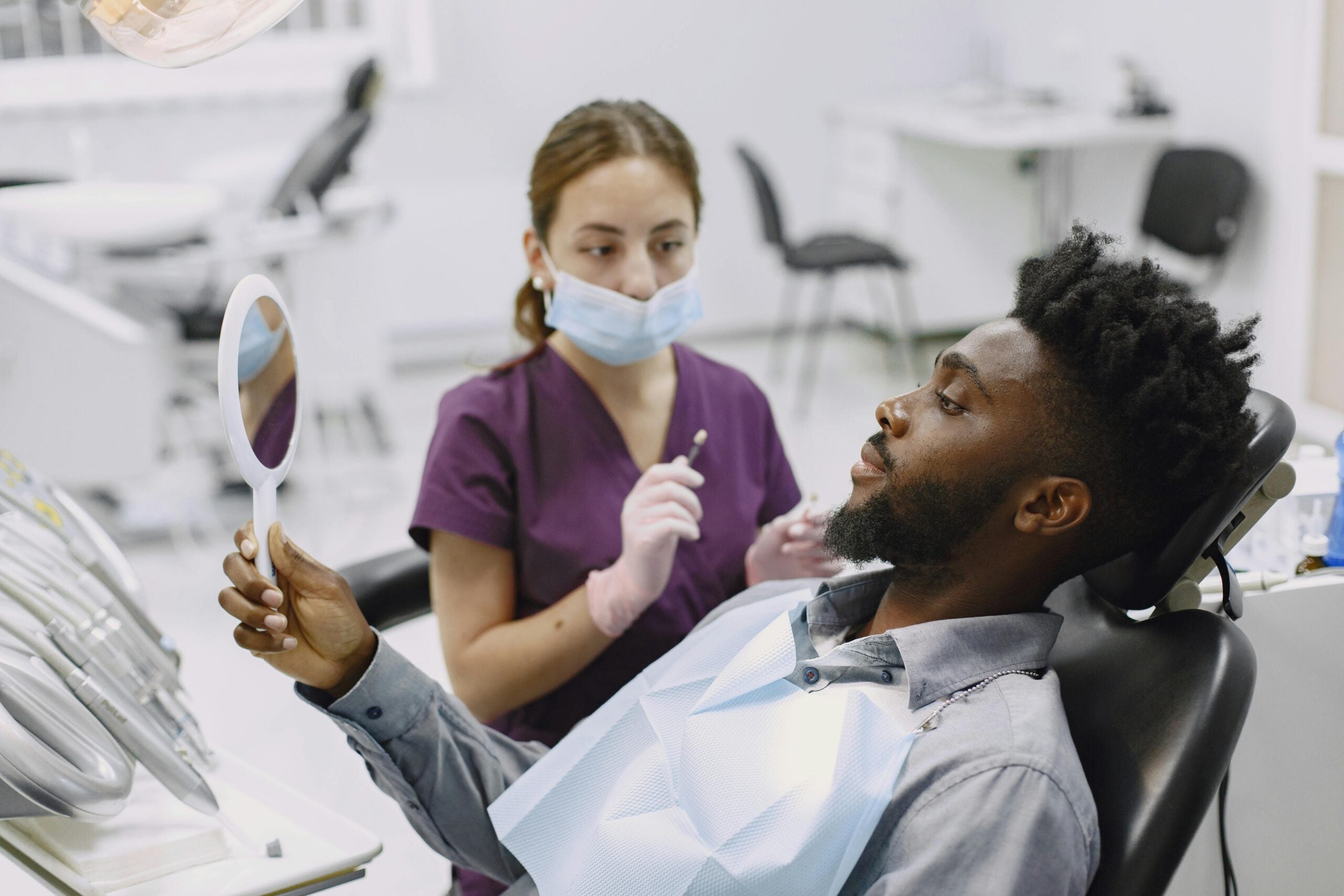
Patient Experiences and Testimonials
Most patients describe the first few days after PRF or PRP as calmer and easier. They often report less bleeding on day one, a steadier clot, and gentler soft tissue healing. Some patients feel only a small difference, which is normal, since results depend on the procedure and the body’s healing response.
Day two after an extraction, chewing soft foods feels easier. With PRF, many notice the site feels protected, like a soft cushion over the wound. That membrane gradually blends into the healing tissue over several days. With PRP, there is usually nothing to feel at the site, since it is a liquid used during surgery. A brief blood draw is part of both options; a mild bruise or soreness at the draw site can occur.
Early comfort is the most common theme. Patients often change gauze fewer times, see less oozing, and return to gentle routines sooner. Swelling still follows its normal arc, typically peaking around day two, but some patients say it feels more manageable. When sutures are used, they hold the area steady while the clot organizes. Good home care, as directed for your case, still matters most for a smooth recovery.
Not everyone has the same experience. Larger defects, thin tissue, smoking, or health conditions like diabetes can blunt the effect. Technique also matters, so two people can have different results even with similar procedures. PRF may feel slightly bulky right after placement, then soften as it integrates; PRP goes unnoticed because it disperses through the site. A light metallic taste from minor bleeding is common on day one and fades as the clot matures.
If you follow prf prp in dentistry evidence, you will see studies focus on patient pain scores, comfort, and early soft tissue closure. That lines up with what patients report in daily life: steadier early healing and easier hygiene in the first week. These tools assist your body, they do not replace it. If you have concerns during recovery, reach out to your dental team for guidance.
Takeaway: expect a steadier early phase, not a dramatic change.
Future Directions in PRF/PRP Research
Research is moving toward clearer rules and stronger trials so we know when PRF or PRP truly helps. Future studies will compare PRF and PRP head-to-head, use standard protocols, and track results that matter to patients, like comfort and function. Researchers also aim to measure long-term bone and gum changes, not just early healing. The goal is simple, match the right material to the right case.
A patient with diabetes asks whether PRF will improve implant healing. To answer well, studies need to sort results by health factors, like smoking, blood sugar control, and medications. They also need to report what was actually placed, including platelet and white cell levels, clot density, and timing. Better imaging and measurements, such as bone volume, soft tissue thickness, and early blood flow at the site, will make findings more useful in daily practice.
Another focus is how these materials are made and handled at the chair. Consistent blood draw volumes, spin settings, and placement steps should be reported in detail. Researchers are testing gentler spin methods and injectable forms that may fit tight spaces. There is interest in small, cell-made packets called extracellular vesicles and how they might carry helpful signals. Safety, sterility, and simple workflows remain the foundation, since the process happens during an active procedure.
Finally, trials should reflect real clinics. That means multicenter studies, clear pain and quality-of-life measures, and follow-up beyond a few weeks. Registries can track results across many cases and help identify who benefits most. Cost and access matter too, since a quick, safe blood draw should not slow care. For those following prf prp in dentistry evidence, expect clearer guidance on which cases gain most and how to tailor use to the site and the patient.
Takeaway: better-designed studies will help clinicians use PRF and PRP more precisely.
Conclusion: Evaluating the Hype
PRF and PRP are helpful, but they are not magic. They tend to improve the first days of healing by protecting the clot and calming tissues, especially in small or moderate procedures. The gains are usually modest, and they depend on careful technique and the biology of the site. In short, the hype is partly earned, but expectations should stay realistic.
Think practical value. When a wound needs stable coverage or a gentle scaffold, PRF often fits. When a site can hold a liquid boost within a contained space, PRP may be chosen. These concentrates work best where early clot protection and blood supply matter most, such as fresh sockets, soft tissue closures, and small defects. They add little when there is poor stability, active infection, or large missing bone volume that requires structural grafting.
Now weigh benefits against limits. Patients often notice easier hygiene, steadier clotting, and less tenderness in the first week. On day one after a premolar extraction, a protected site often feels calmer. However, long-term bone shape, implant success, and bite function still rely on sound surgery, healthy blood flow, and good home care. Habits and health conditions, like smoking or diabetes, can blunt any added effect. If you follow prf prp in dentistry evidence, the pattern is consistent: small early gains are possible, yet quality and uniformity of studies vary, so results are not guaranteed across all procedures.
How should you decide? Match the material to the job. Choose PRF when you need a simple, easy-to-handle cover that stays put. Choose PRP when a quick, contained biologic rinse makes sense. Ask what problem the concentrate will solve at your site, and what other steps, such as suturing or grafting, will support the plan. Clear goals, clean fields, and stable closure matter more than any single adjunct.
If you are considering these options, discuss your health history, healing goals, and the specific procedure with your dentist. For practical planning and timing, check our current hours. Takeaway: use PRF or PRP when they solve a clear, early-healing problem.
Frequently Asked Questions
Here are quick answers to common questions people have about PRF/PRP: Hype or Helpful? in Glendale, AZ.
- What are the primary differences between PRF and PRP in dentistry?
PRF (platelet-rich fibrin) and PRP (platelet-rich plasma) both use your blood to aid healing, but they differ in preparation and function. PRP is a liquid that quickly releases growth factors when activated, while PRF forms a gel-like clot releasing signals over time. PRF often serves as a protective scaffold for wound coverage, and PRP is used for a rapid boost in cellular signals. The choice depends on whether the procedure benefits more from immediate signals or sustained tissue support.
- How do PRF and PRP support healing in dental surgeries?
In dental surgeries, PRF and PRP enhance healing through concentrated platelets that release growth factors and cytokines. PRF forms a stable matrix, providing a structure for new tissue growth. PRP delivers an early, intense burst of biological signals that can kickstart tissue repair. Their combined effects can lead to quicker tissue closure, reduced early discomfort, and better bleeding control, making the recovery phase smoother and more comfortable.
- What are the potential benefits of using PRF or PRP after a tooth extraction?
When used after tooth extractions, PRF and PRP can provide several benefits. They can help stabilize the clot, protecting the surgical site from early breakdown. This stability can lead to less bleeding, lowered risk of complications like dry socket, and more organized tissue growth. Patients may experience improved comfort and find it easier to maintain oral hygiene during the initial healing. These advantages help ensure a better recovery process.
- Why are standardized protocols important in PRF and PRP applications?
Standardized protocols are crucial for the effective use of PRF and PRP because they ensure consistent quality and outcomes. Variations in techniques, like spin speed and preparation methods, can lead to different product characteristics, affecting effectiveness. Standardization helps clinicians and researchers compare results, refine techniques, and improve the reliability of these treatments across various dental procedures, ensuring safer and more predictable outcomes for patients.
- Can PRF and PRP replace traditional dental treatments?
PRF and PRP are not replacements for traditional dental treatments but act as adjuncts. They do not substitute for careful surgical technique, suturing, or good oral home care. Their role is to support early healing and comfort by providing a biologic scaffold or rapid cellular signaling. While beneficial, their effects are modest and best paired with comprehensive dental care plans for optimal results in procedures like extractions or implants.
References
- [1] PRF and PRP in Dentistry: An Umbrella Review. (2025) — PubMed:40364255 / DOI: 10.3390/jcm14093224
- [2] Platelet-Rich Plasma and Platelet-Rich Fibrin in Endodontics: A Scoping Review. (2025) — PubMed:40564941 / DOI: 10.3390/ijms26125479
- [3] Use of platelet concentrates in oral and maxillofacial surgery: an overview. (2017) — PubMed:27669885 / DOI: 10.1080/00016357.2016.1236985
- [4] Extended platelet-rich fibrin. (2024) — PubMed:37986559 / DOI: 10.1111/prd.12537
- [5] Effectiveness of platelet-rich fibrin or platelet-rich plasma in mandibular fracture management: A systematic review and meta-analysis. (2025) — PubMed:40915418 / DOI: 10.1016/j.jormas.2025.102544
- [6] The evolution of three generations of platelet concentrates products: a leap from classical formulations to the era of extracellular vesicles. (2025) — PubMed:40851809 / DOI: 10.3389/fbioe.2025.1628565