What is Socket Preservation?
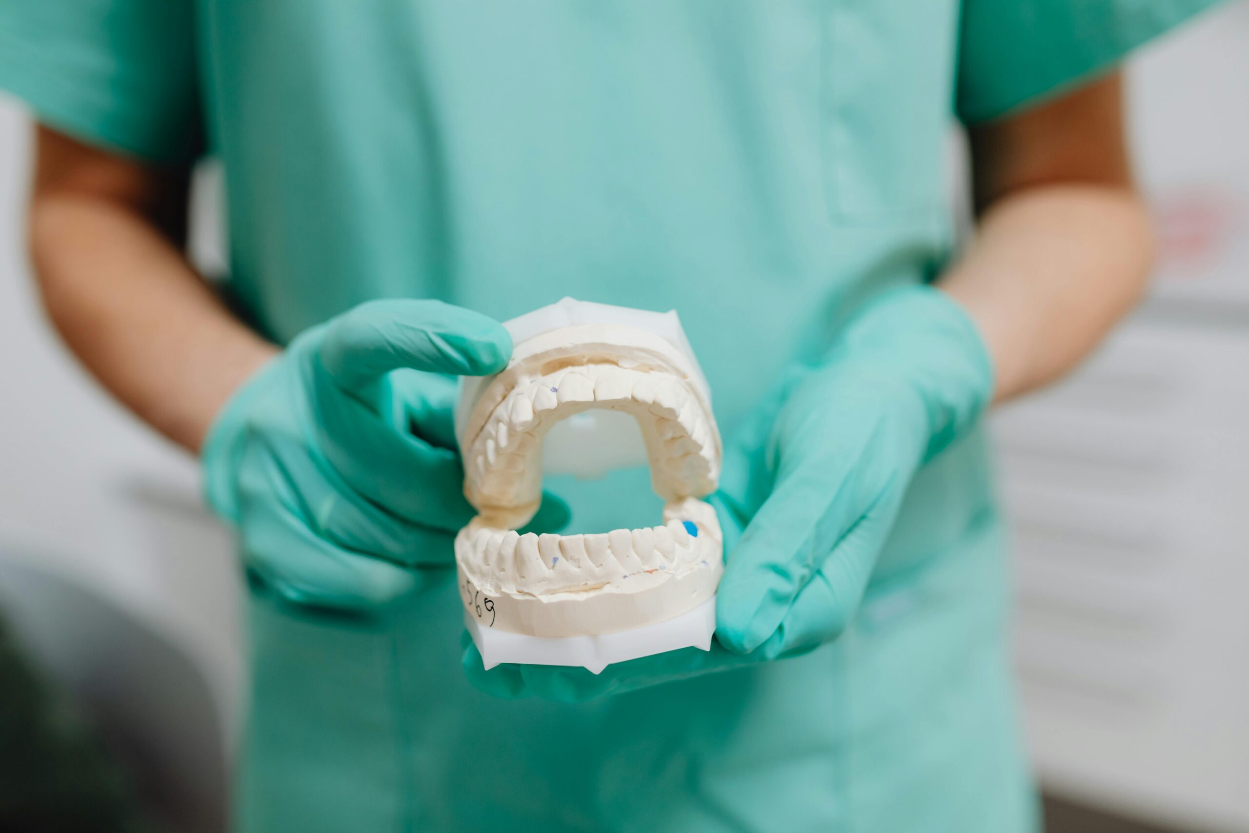
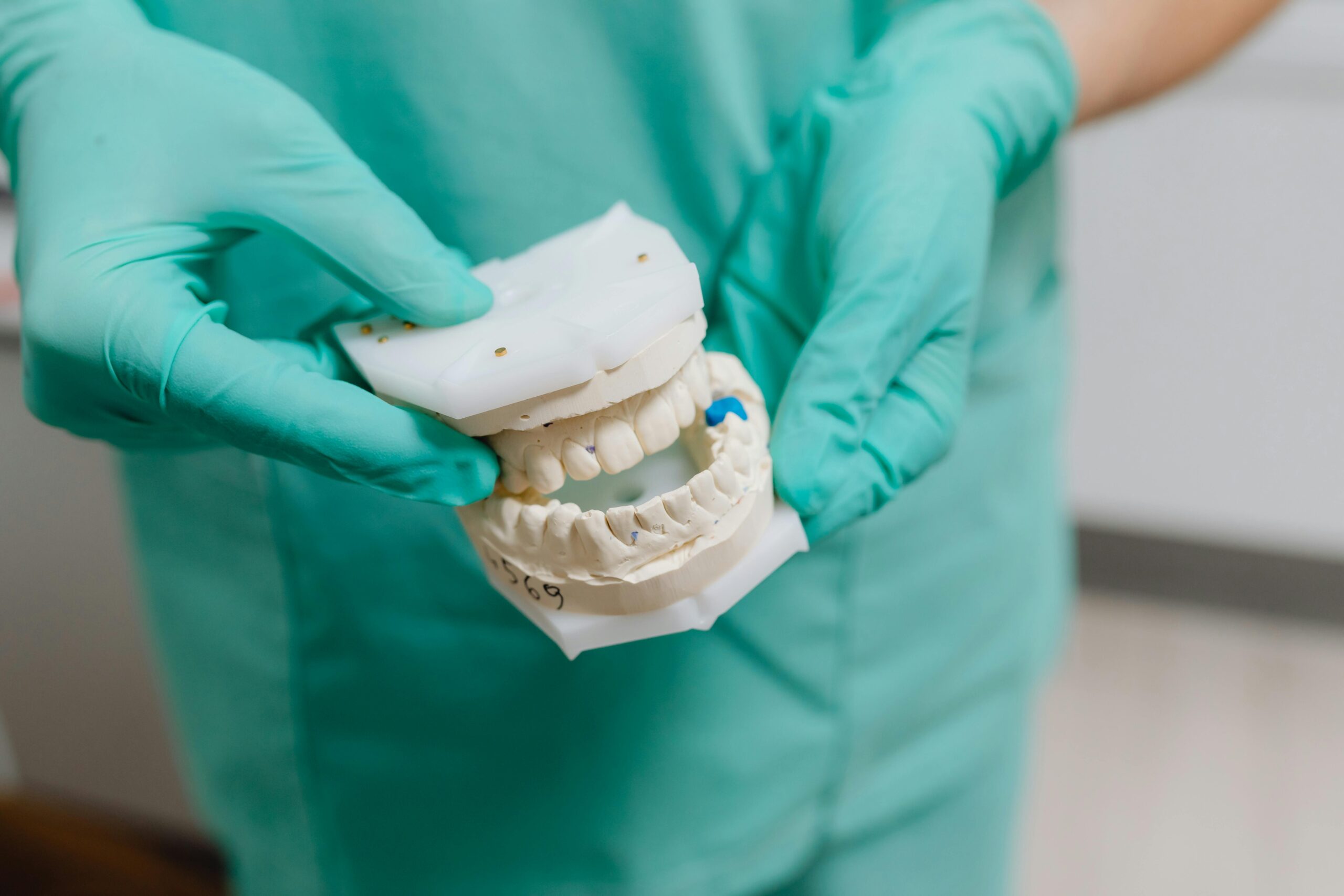
Socket preservation is a minor procedure performed at the time of tooth extraction to help maintain the bone where the tooth once sat. After the tooth is removed, the empty socket is gently filled with bone graft particles and often covered with a thin membrane, then closed with small stitches. This supports the blood clot, helps the area heal with more bone, and maintains the ridge shape for future options such as an implant or bridge.
Why is this done? After an extraction, the body naturally reshapes and resorbs the thin outer bone, which can leave a dip in the gumline and less bone for tooth replacement. The graft acts as a scaffold your body remodels into your own bone over several months (commonly 3–6), while the membrane protects the site during early healing. Materials can be human donor, animal-derived, or synthetic; the choice depends on the site, infection risk, and your goals. It is not needed in every case, but it is especially helpful when an implant is planned, the front teeth are involved, or the socket walls are thin. Here is the socket preservation bone graft explained in plain language: it buys time and shape so you have more options later.
- Bite gently on the gauze placed by your dentist to control normal oozing.
- Do not disturb the site: avoid straws, vigorous rinsing, spitting, or smoking.
- Use a cold compress on the cheek in short intervals the first day to reduce swelling.
- Choose soft foods, chew on the other side, and rest with your head slightly elevated.
Clean nearby teeth gently; avoid brushing directly on the graft for 24 hours. If pain, persistent bleeding, unusual swelling, or the membrane feels loose, contact the office during business hours for an in-person check.
Benefits of Socket Preservation
Socket preservation helps maintain the natural width and height of the jawbone after a tooth is removed. By supporting the extraction site as it heals, it reduces the rapid bone shrinkage that typically follows an extraction. This keeps options open for future tooth replacement, often making dental implant or bridge placement more predictable and esthetic.
- Helps preserve ridge shape and facial support
- Supports gum contours for a more natural look
- May reduce the need for larger grafts later
- Improves planning and positioning for future implants
Can limit shifting of nearby teeth after extraction
If you want the socket preservation bone graft explained simply: a small graft material is placed in the socket to stabilize the ridge while your body forms new bone. To support healing at home, avoid smoking, don’t disturb the site, choose soft foods, and use gentle saltwater rinses after the first day if advised. If you notice worsening pain, swelling, fever, or persistent bleeding, contact a dentist promptly during business hours for an in-person evaluation.
Materials Used for Bone Grafting
Bone grafts placed at the time of tooth removal act as a temporary scaffold, preserving space and supporting your own bone as it heals. Options vary by source and how quickly they dissolve, but all are selected for biocompatibility and safety. Consider this socket preservation bone graft explained through the materials commonly used and why one may be chosen for your situation.
- Autograft (your own bone): Contains living cells and natural growth factors; excellent integration, but requires a small donor site.
- Allograft (donor human bone): Thoroughly processed and sterilized; commonly used, with predictable remodeling into your bone.
- Xenograft (animal-derived mineral): Slow-resorbing scaffold that helps maintain volume in areas at higher risk of collapse.
- Alloplast (synthetic materials): Calcium phosphate or similar minerals; avoids human/animal sources, with resorption rates tailored to the case.
Barrier membranes and biologics: Thin resorbable membranes keep gum tissue out of the graft while it matures; your own blood-derived concentrates may be used to support early healing.
Your dentist chooses materials based on anatomy, timing for any future implant, medical history, and desired healing speed. After the procedure, avoid disturbing the site (no vigorous rinsing, spitting, or touching), follow any prescribed medication plan, and stick to a soft diet until advised otherwise. If you notice worsening pain, persistent bleeding, or a large amount of sandy graft particles coming out, contact the office during business hours so we can examine the area and guide next steps.
Healing Timeline After Extraction
After an extraction, healing follows a predictable arc for most patients. In the first 24 hours a protective blood clot forms; swelling often peaks around days 2–3 and then subsides through the first week. If graft particles and a small membrane were placed, it’s normal to notice a slight “gritty” feel early on—soft tissue typically closes in 2–3 weeks while the graft integrates over 8–12 weeks, with full bone remodeling taking about 3–4 months. You’ll often hear socket preservation bone graft explained as a way to maintain the ridge’s shape for future treatment.
- First 24 hours: Bite gently on gauze as instructed to control oozing; rest, keep your head elevated, and avoid smoking, straws, or vigorous rinsing.
- 48–72 hours: Expect peak swelling and possible minor bruising; use cold compresses 10 minutes on/off during day one and continue to sleep with your head elevated.
- Day 2 onward: Rinse gently with warm saltwater 2–3 times daily; brush and floss other teeth as usual, taking care near the site.
- First week: Favor a soft diet and chew on the opposite side; avoid probing the socket or dislodging any membrane or sutures.
1–2 weeks: Many sutures are removed; light exercise can resume if comfortable; continue gentle cleaning around the area.
Please contact the office during business hours promptly if pain worsens after day 3, swelling rapidly increases, fever develops, you notice pus or persistent bleeding, or if a covering membrane becomes loose.
Procedure Overview for Socket Preservation
Socket preservation is a brief add-on procedure performed immediately after a tooth is removed to help maintain the natural contour of the jawbone. Here is socket preservation bone graft explained in simple terms: a small amount of biocompatible graft is placed into the empty socket, covered, and secured so your body can replace it with new bone over time. This helps keep options open for future restorations and can make the area easier to clean and restore.
- Evaluation and planning, often with an X‑ray
- Local anesthesia for comfort
- Gentle, atraumatic tooth removal
- Thorough cleaning of the socket
Placement of graft material to fill the socket
Covering with a membrane or collagen plug when indicated
Sutures to secure the site and protect healing
After you leave, expect mild oozing the first day and some swelling for 48–72 hours. At home, rest, keep the head elevated, bite gently on the provided gauze until bleeding slows, avoid vigorous rinsing or straws for 24 hours, use a cold compress in short intervals the first day, choose a soft diet, and consider over‑the‑counter pain relievers as directed on the label. Starting the next day, gentle warm salt‑water rinses can help keep the area clean. A follow‑up visit is typically scheduled to check healing and, if used, remove sutures. If you notice persistent bleeding, fever, or increasing pain, please contact the office promptly during business hours for in‑person care.
Managing Discomfort and Recovery
Here, you’ll find socket preservation bone graft explained in practical terms: most people experience manageable soreness and swelling that improve over several days. Comfort comes from protecting the graft, keeping the area clean, and giving your body time to heal. The steps below can help you feel better while you recover and know when to contact our office. Some bruising is normal and typically peaks around day two or three before fading.
- Rest the day of the procedure and keep your head slightly elevated to limit swelling; avoid heavy activity for 24 hours.
- Apply a cold compress to the cheek 15 minutes on, 15 off during the first 24–48 hours.
- Bite gently on gauze as directed until oozing slows; avoid vigorous spitting or using straws.
- Choose soft, cool foods and plenty of fluids; avoid smoking or vaping while you heal.
Starting 24 hours later, rinse gently with warm salt water and brush carefully around the area without disturbing the graft.
Over-the-counter pain relievers may help; use only what you tolerate and follow label directions. Call our office during business hours if bleeding won’t slow, pain worsens after day 3, or you notice fever, increasing swelling, foul taste, or drainage.
Next Steps After Socket Preservation
With the socket preservation bone graft explained at your visit, the immediate goals are to protect the graft, stay comfortable, and set up a smooth healing plan. Rest the day of the procedure and avoid disturbing the area. Call our office during business hours with any questions, and plan to attend your follow-up so we can confirm the site is healing as expected.
- Bite gently on gauze for 30–45 minutes; slight oozing is normal. Replace as instructed if needed.
- Avoid spitting, rinsing forcefully, or using straws for 24 hours; do not smoke or vape.
- Use a cold compress on-and-off (15 minutes each) for the first day to limit swelling.
- Choose soft, cool foods; chew on the opposite side and stay hydrated.
Brush other teeth as usual; near the site, brush carefully without touching sutures or any protective membrane. After 24 hours, start gentle saltwater rinses after meals.
Take pain relievers only as directed by your dentist or per the label.
Expect mild swelling that peaks around 48–72 hours and gradually improves. You may notice a few tiny granules in your saliva; don’t touch the area. Please contact the office during business hours if you have persistent bleeding, increasing pain after day three, expanding swelling, fever, bad taste or discharge, or if a membrane or suture loosens. We’ll schedule in-person checks to monitor healing and discuss when it’s safe to return to normal brushing and diet over the grafted site.
Long-Term Care for Grafted Sites
In the months after a graft, the goal is to protect the surgical site while your body gradually replaces the graft with your own bone. Consistent, gentle home care and routine check-ins help maintain the ridge and reduce complications. Think of this as your socket preservation bone graft explained in plain terms for day-to-day care.
- Brush the rest of your teeth twice daily with a soft brush; near the grafted spot, clean carefully and avoid direct scrubbing until your dentist clears it.
- After 24 hours, rinse gently with lukewarm saltwater 2–3 times a day; avoid vigorous swishing and strong mouthwashes unless instructed.
- Do not pull on your cheek or lip, and avoid straws, forceful spitting, smoking, and vaping.
- Choose soft, cool foods for the first few days, then return to a balanced diet with adequate protein; stay well hydrated.
Do not disturb any dressing or membrane; a few sandy granules early on can be normal, but call us if you notice ongoing material loss or the cover lifting.
Keep plaque under control: floss adjacent teeth carefully and avoid poking the grafted area.
Contact our office promptly during business hours for heavy bleeding, worsening pain after day two, increasing swelling, fever, or a bad taste/odor.
Gum tissue often feels settled within a couple of weeks, but bone remodeling continues for several months. Mild stiffness or brief twinges can occur as tissues adapt. Attend all scheduled reviews so your dentist can confirm stability and advise when it’s safe to move to the next step of your treatment. If anything feels off, we want to see you—please reach out during business hours for timely in-person care.
Frequently Asked Questions
Here are quick answers to common questions people have about Socket Preservation 101: Saving Bone After Extraction in Glendale, AZ.
- What is socket preservation and why is it important?
Socket preservation is a procedure performed after a tooth extraction to help maintain the bone that supported the tooth. It involves placing bone graft material in the empty socket to support healing and preserve the ridge shape. This is particularly important for future tooth replacement options like dental implants or bridges, as it prevents the natural bone loss that typically follows an extraction.
- How does socket preservation help with future dental implants?
Socket preservation reduces the rapid bone shrinkage often seen after an extraction. By maintaining the width and height of the jawbone, it makes dental implant placement more predictable. This ensures better esthetics and functional outcomes, as implants require sufficient bone for support.
- What materials are used for socket preservation bone grafts?
Bone graft materials vary and include autografts (your own bone), allografts (processed human donor bone), xenografts (animal-derived mineral), and alloplasts (synthetic materials). Each type has unique properties regarding integration and resorption rates, so your dentist will choose based on your specific needs and medical history.
- What should I expect during the healing process after socket preservation?
Initially, you’ll experience mild oozing and swelling. Over the first week, swelling should decrease. The graft material integrates with your bone over 8–12 weeks, with full bone remodeling taking about 3–4 months. Follow your dentist’s care instructions carefully to ensure proper healing.
- How can I manage discomfort after a socket preservation procedure?
Rest and keep your head elevated to minimize swelling. Use cold compresses the first couple of days, and take any prescribed or over-the-counter pain relievers as directed. Stick to a soft diet and avoid disturbing the site by not using straws or spitting vigorously. Gentle oral hygiene is important but avoid direct contact with the graft area.
- Why might socket preservation not be needed in every case?
Socket preservation is most beneficial when future dental restorations like implants are planned, where front teeth are involved, or where socket walls are thin. In cases where these factors aren’t present, or when immediate bone resorption isn’t a concern, the procedure might not be necessary.
- What activities should I avoid after socket preservation to ensure healing?
Avoid smoking, using straws, vigorous rinsing, and spitting as these activities can dislodge the healing clot and graft material. Stick to soft foods and stay hydrated, but avoid chewing on the graft side until cleared. Smoking, in particular, can significantly hinder healing.
- What should I do if I notice unusual symptoms after the procedure?
If you experience persistent bleeding, increasing pain after day three, swelling, fever, or loosening of the graft membrane, contact your dentist promptly during business hours. These could be signs of complications that require professional assessment.
Medical sources (PubMed)
- Mularczyk C, et al. Otolaryngol Clin North Am. 2024. “Maxillary Sinus Anatomy and Physiology.”. PMID: 39142996 / DOI: 10.1016/j.otc.2024.07.004
- Camps-Font O, et al. Int J Oral Maxillofac Surg. 2024. “Antibiotic prophylaxis in the prevention of dry socket and surgical site infection after lower third molar extraction: a network meta-analysis.”. PMID: 37612199 / DOI: 10.1016/j.ijom.2023.08.001
- Rolek A, et al. Wiad Lek. 2024. “Comprehensive management of pericoronitis in lower third molars: extraction, operculectomy, and coronectomy approaches.”. PMID: 39241154 / DOI: 10.36740/WLek202407129
- Garaycochea O, et al. Curr Allergy Asthma Rep. 2023. “Pheno-Endotyping Antrochoanal Nasal Polyposis.”. PMID: 36773125 / DOI: 10.1007/s11882-023-01066-1
- Chung SY, et al. Curr Opin Otolaryngol Head Neck Surg. 2023. “Tips and tricks for management of the dysfunctional maxillary sinus.”. PMID: 36484283 / DOI: 10.1097/MOO.0000000000000860
- Steel BJ, et al. Br J Oral Maxillofac Surg. 2022. “Current thinking in lower third molar surgery.”. PMID: 34728107 / DOI: 10.1016/j.bjoms.2021.06.016

