Importance of CBCT in Dental Planning
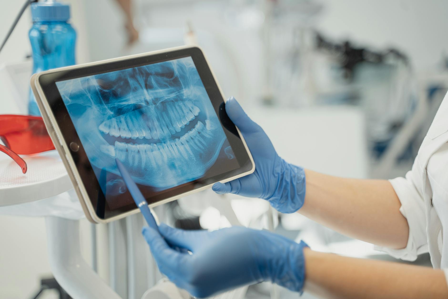
Cone-beam computed tomography (CBCT) gives a true 3D view of your jaws and teeth, which standard dental X-rays cannot provide. In dental planning, this detail helps map bone shape and thickness, locate nerves and sinuses, and spot hidden problems so the plan fits your exact anatomy. This is especially important for complex care like full-arch implant restorations and surgical extractions. CBCT data also supports accurate, guide-assisted surgery.
For full-arch cases, planning starts with the final teeth in mind. Using cbct for full-arch implants lets the team position implants to support a stable bite, clear speech, and easy cleaning, while avoiding vital structures. The 3D scan shows whether grafting or a sinus lift is needed, how many implants are appropriate, and the angulation needed for a prosthetic design that lasts. When a patient is a candidate, CBCT helps plan immediate or staged loading strategies and the fabrication of precise surgical guides for options such as All-on-4 implant dentures.
- Mapping the mandibular canal and submandibular fossa reduces the risk of nerve injury in the lower jaw [1].
- Visualizing the nasopalatine (incisive) canal helps avoid this structure during anterior maxillary implant placement [2].
- Locating the maxillary sinuses and floor thickness informs sinus lift decisions.
- Measuring bone volume and quality guides whether grafting or shorter, angled implants are better.
- Screening for cysts, infections, or root fragments prevents surprises on surgery day.
- Checking vertical space helps the lab design a strong, cleansable full-arch prosthesis.
Because CBCT ties surgical and restorative goals together, it improves communication among the surgeon, restorative dentist, and lab. The scan can be merged with digital impressions to design a prosthetic-driven plan and to fabricate a custom guide that transfers the plan to the mouth with high precision. These same planning steps also apply to removable options like snap-in implant dentures, where implant position and angulation are key for retention and comfort.
Understanding Full-Arch Implants
Full-arch implants replace all the teeth in one jaw with a prosthesis that is anchored to several dental implants. The goal is to restore chewing, speech, and appearance with a design that feels stable and is simple to clean. Depending on your needs, the final prosthesis can be fixed (removed only by the dentist) or removable (snaps in and out).
Think of each implant as a new “root” in the bone. After the implants are placed, connectors (abutments) support either a one-piece fixed bridge or an implant denture that engages attachments. Fixed options feel the most tooth-like; removable designs can be easier to clean at home. Your team will match the approach to your jaw anatomy, bite, dexterity, and hygiene habits.
Several factors guide whether you are a good candidate and how many implants are advisable: bone volume and quality, the way your teeth fit together, bite forces, parafunction (like clenching or grinding), gum and medical health, and your ability to maintain daily hygiene. Managing occlusion and overall biomechanics is central to long-term success, because overload can lead to screw loosening, fractures, or bone loss if not addressed in the plan [3].
Timing of the teeth also varies. In some cases, a provisional full-arch bridge can be attached the same day (“immediate loading”). This requires strong primary stability at placement and careful control of the bite on the temporary prosthesis; when those prerequisites are not met, a staged approach allows healing before loading [4].
Planning is “prosthetic-driven,” meaning implants are placed to support the final teeth, not the other way around. Records include digital impressions, jaw relation measures, and imaging—often cbct for full-arch implants—to map bone and vital structures so the prosthesis is strong, cleansable, and comfortable. Comfort during longer appointments can be supported with options such as oral sedation for dental treatment, when appropriate. Like natural teeth, full-arch prostheses need ongoing maintenance: daily cleaning, periodic professional cleanings, and checkups to monitor screws, acrylic or ceramic surfaces, and the tissues underneath. Full-arch implant–retained prosthetics are an established option in general dental practice when planned and maintained carefully [5].
Anatomical Considerations in Implant Placement
Successful implant placement depends on knowing the exact shape of your jaws, where nerves and sinuses travel, and how much space the final teeth need. The plan must protect vital structures while placing implants at angles and depths that support a strong, easy-to-clean prosthesis. Using cbct for full-arch implants gives a true 3D map of these details so surgery and the final bridge line up.
In the upper jaw, the maxillary sinuses limit where posterior implants can go and whether grafting or angled strategies are better. In the front of the maxilla, the nasopalatine (incisive) canal can vary in size and position; identifying it on CBCT helps avoid that structure during implant placement and reduces the chance of symptoms in the palate region [2]. Thin facial bone, ridge undercuts, and the smile line also guide implant positions so the prosthesis has proper support and natural-looking transitions.
In the lower jaw, the inferior alveolar nerve within the mandibular canal, the mental foramen and its anterior loop, and the submandibular fossa concavity are key landmarks. CBCT helps measure how thick the bone is on the cheek and tongue sides, check vertical height above the nerve, and spot lingual undercuts so implants stay in safe bone. Space for the prosthesis matters too; the vertical room between jaws must allow strong materials, hygiene access, and comfortable speech.
When the posterior maxilla has limited bone because of sinus pneumatization, alternative anchorage such as pterygoid implants may be considered. These approaches depend on the pterygo-maxillary anatomy, and classification systems help guide safe placement paths in this region [6]. Even with advanced options, the goal remains the same: stable support while avoiding nearby vessels, nerves, and sinus walls.
Before surgery, the team reviews CBCT alongside digital impressions to design a prosthetic-driven plan that respects these anatomical limits. This includes mapping nerves and canals, measuring bone volume and density, and confirming restorative space so the final bridge is strong and cleansable [7]. With this roadmap, the surgical guide and the restoration are coordinated, lowering surprises and helping treatment proceed smoothly.
How CBCT Enhances Treatment Accuracy
CBCT enhances accuracy by giving a true 3D map of your jaws so the plan can be matched to your exact anatomy. For full-arch care, the team can plan implant positions on the computer, then transfer that plan to your mouth with a surgical guide or real-time navigation. This reduces surprises, helps avoid nerves and sinuses, and supports a bridge that fits your bite and is easy to clean. Using cbct for full-arch implants helps align the surgical steps with the final teeth.
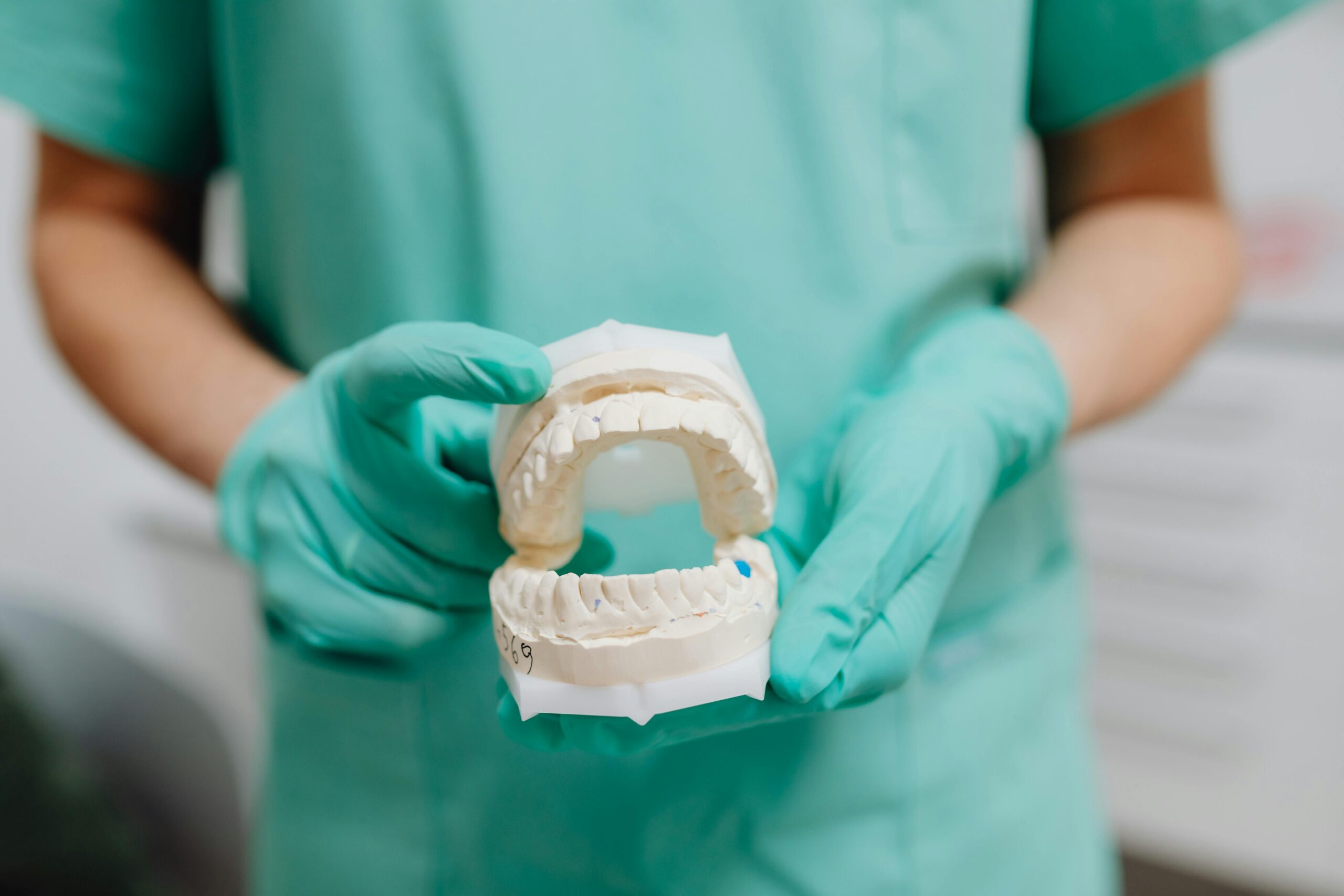
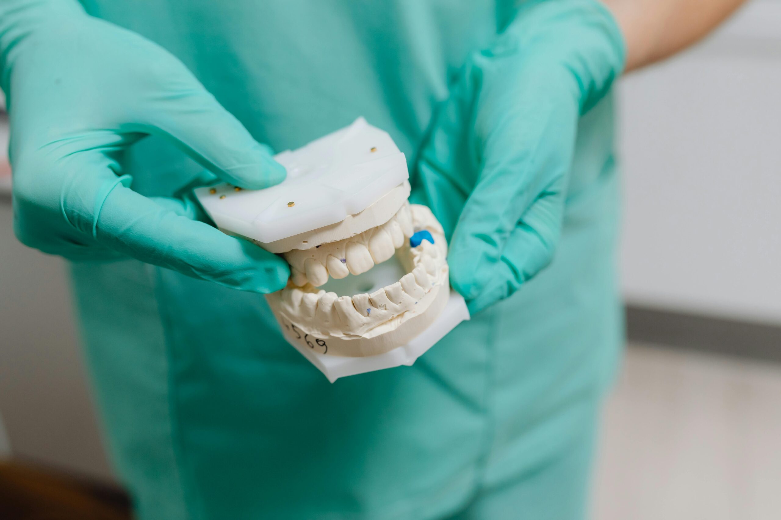
The process starts by merging the CBCT scan with digital impressions of your gums and teeth. This creates a single model that shows bone, soft tissues, and the planned teeth together. The team sets safe distances from vital structures, chooses implant sizes, and adjusts angulation so screw channels and hygiene access are in the right places. They also check vertical room for the prosthesis, so there is enough space for strong materials and clear speech.
Next, a custom guide is made to “lock in” the plan. Depending on your case, the guide may rest on teeth, gums, or bone, and small anchor pins can be used to keep it steady. A snug fit is key because less guide movement means closer match to the plan. Drill sleeves and stop lengths help control depth and angle. Some offices use dynamic navigation, which tracks the handpiece against the CBCT in real time; whether static or dynamic, careful calibration and fit checks are what protect accuracy.
On surgery day, the team verifies the guide seats fully, checks access for the handpiece, and confirms that the planned path is clear. During drilling, they maintain steady support and irrigation to avoid heat. After placement, the implants are compared with the plan, and a provisional bridge is adjusted to keep bite forces even while you heal. These steps lead to a better match between planned and placed implant positions, safer distances from vital structures, and a prosthesis that functions well. CBCT adds targeted, 3D information that improves decisions at every step, from planning to placement to follow-up.
Airway Assessment with CBCT Imaging
CBCT can show a 3D view of your nasal passages and throat (the upper airway) along with the jaws and teeth. While it does not diagnose sleep apnea, it helps the team see airway shape and available space that might matter during planning. Using cbct for full-arch implants also lets us confirm there is enough room for the tongue and for a prosthesis that does not crowd the throat.
On a CBCT, we review the nasal cavity, sinus openings, the back of the nose (nasopharynx), and the space behind the tongue (oropharynx). We can measure airway volume and the tightest cross-sectional area, and relate those findings to jaw shape and position [8]. These images provide context: if the airway looks narrow or the nose appears blocked, we can design the prosthesis to avoid bulky contours and note when medical evaluation could be useful.
For full-arch planning, airway review ties directly to prosthetic space. In the upper jaw, a fixed bridge does not cover the palate, which can help speech and reduce bulk. In the lower jaw, we shape the tongue-side contours to be smooth and cleansable without pushing into the tongue space. CBCT also helps confirm vertical room between the jaws so the teeth, framework, and hygiene access fit without encroaching on the airway corridor.
It is important to set expectations: a CBCT is a snapshot taken in an upright, awake state, and breathing and head posture can change airway size from moment to moment. Even so, CBCT is reliable for showing anatomical changes after jaw surgery, which reminds us that jaw position can influence airway dimensions [9][10]. Implants do not move the jaws, but they do support new tooth positions and vertical dimension; we plan these carefully so the final teeth function well and feel natural while respecting tongue and airway space. If the scan suggests notable crowding or nasal blockage, we can coordinate with your physician or a sleep specialist for further testing when appropriate.
Minimizing Surgical Risks with CBCT
CBCT reduces surgical risks by showing a clear 3D map of your jaws before any work begins. It helps the team see nerves, sinuses, and areas where the bone curves or thins, so implant paths can be planned to stay in safe zones. This planning lowers the chance of surprises and supports careful, guide-assisted surgery when needed.
In the lower jaw, CBCT can reveal the exact shape and thickness of the inner (tongue-side) bone. Seeing these concavities ahead of time helps avoid lingual plate perforation and related bleeding during implant drilling or extraction in the posterior mandible [11]. CBCT also shows the mental foramen and any forward “loop” of the nerve, which helps the surgeon choose safe implant lengths and entry points to reduce the risk of numbness or pain after surgery [12].
In the upper jaw, CBCT clearly outlines the maxillary sinuses and nearby blood vessel pathways, such as the greater palatine canal. Knowing the sinus floor height and palatal canal course helps plan implant positions and sinus-lift approaches that avoid membrane tears and vessel injury, which can reduce complications like sinus perforation or bleeding [13]. This same 3D detail helps check bone thickness at the nasal floor and front of the maxilla, where roots, canals, or thin plates may limit safe implant angulation.
Beyond mapping anatomy, CBCT supports safer workflows. The scan can be merged with digital impressions to design a custom surgical guide, which helps control implant angle and depth and reduces “drift” from the plan. During the procedure, steady guide support and irrigation help keep drills on track and reduce heat. After placement, the team can verify implant positions against the plan and adjust the temporary bridge to keep biting forces even while you heal. When used thoughtfully—especially in cbct for full-arch implants—this step-by-step approach lowers the chances of nerve injury, sinus complications, cortical perforations, and misaligned implants, and it improves the odds of a strong, cleansable final prosthesis.
Interpreting CBCT Data for Implants
Interpreting CBCT data means turning a 3D scan into a clear, step-by-step implant plan. We measure bone, find important structures like nerves and sinuses, and relate everything to the planned teeth and bite. For full-arch cases, we also check space for the bridge and hygiene access, so the plan supports strong, easy-to-clean teeth. Using cbct for full-arch implants helps keep surgery and the final bridge aligned.
The team reviews the scan in thin “slices,” moving through the jaws from different angles to see true bone height and width at each planned implant site. We measure from the crest to nearby limits (such as a nerve canal or sinus floor), check for undercuts, and look for thin spots in the outer bone that might need a different angle or grafting. CBCT can reveal facial (cheek-side) bone defects that matter for immediate implant plans, and even very low-dose protocols have been studied for this purpose in ex vivo models [14]. These details guide implant length, diameter, and position before any drilling begins.
Next, we trace the anatomy that must be protected. This includes the inferior alveolar nerve and mental foramen in the lower jaw, the sinus floor and nasal spaces in the upper jaw, and canals such as the incisive and lingual foramina. CBCT-based analyses have mapped the lingual foramen and related variations, which helps clinicians identify risk areas in the front of the lower jaw and plan safe entry paths [15]. We also confirm there is enough vertical room for the final prosthesis materials and for daily cleaning tools to fit comfortably.
Image quality and limitations are part of the read. Motion, metal fillings or old crowns, and very low-dose settings can create streaks or blur that hide edges. Methods to reduce metal artefacts—including deep learning approaches—are being investigated to improve clarity in low-dose dental CBCT scans with high-attenuation materials [16]. Finally, we cross-check the CBCT with digital impressions and a bite record, so the bone picture, planned teeth, and your jaw movements all agree. This careful interpretation narrows uncertainties, supports safe distances from vital structures, and improves the odds of a bridge that fits, functions, and cleans well.
Integrating CBCT into Clinical Practice
CBCT becomes reliable in daily care when it follows a clear, repeatable workflow: capture a good scan, interpret it systematically, merge it with dental records, and use it to guide treatment. For full-arch cases, this ties the surgeon, restorative dentist, and lab together so planning, surgery, and the final teeth stay aligned. Using cbct for full-arch implants helps the team see bone and vital structures, choose implant positions, and design a prosthesis that fits your bite and is easy to clean.
Start with case selection and dose control. Choose a field of view large enough to include both arches and key landmarks (sinuses, nasal floor, mandibular canal) without scanning more than needed. Stabilize the head, remove loose appliances, and set a light bite so jaw position is reproducible. After the scan, do a quick quality check for motion or metal streaks; if artefacts hide anatomy, minor adjustments or a targeted re-scan may be necessary under “as low as reasonably achievable” (ALARA) principles.
Next, merge the CBCT with digital impressions and a bite record to build one model that shows bone, soft tissues, and planned teeth. If a patient has an existing denture, it can be duplicated as a scan appliance to mark tooth position for planning. The team then selects implant sizes, sets safe distances from nerves and sinuses, and confirms vertical room for strong materials and hygiene access. A custom surgical guide is designed and checked on printed models or in the mouth to verify fit before surgery. For longer appointments, comfort can be supported with options such as deep sedation for complex dental procedures when appropriate.
Finally, build habits around interpretation, documentation, and follow‑through. Assign who reads every scan and record key measurements, implant plans, and any incidental findings that may need medical or dental referral. On surgery day, verify the guide seats fully and that access for instruments is clear; after placement, compare implant positions with the plan and adjust the temporary bridge so forces are even. Secure storage of CBCT data and sharing with the lab or referring clinicians should follow privacy rules. For scheduling or coordination questions, check our current hours.
CBCT vs Traditional Imaging Methods
CBCT creates a 3D picture of your jaws, while traditional dental X‑rays (panoramic, periapical, cephalometric) are 2D. For implant planning—especially full-arch cases—3D data shows true bone shape, thickness, and the location of nerves and sinuses. Traditional images remain useful for screening and checking teeth, but CBCT answers depth and distance questions that 2D films cannot. That is why many teams prefer cbct for full-arch implants when precise mapping is needed.
Panoramic X‑rays give a broad overview, but magnification and distortion vary across the image. Periapicals show fine detail around a few teeth, yet they lack depth information and wider context. Cephalometric radiographs help assess jaw relationships, but they do not measure implant sites in three dimensions. CBCT provides undistorted, cross‑sectional views so clinicians can measure from the crest to the mandibular canal or sinus floor, confirm ridge width, and plan safe implant angles. Comparative dental research also shows that CBCT can reveal tooth and jaw morphology more clearly than pantomography, supporting decisions that depend on precise anatomy [17].
In practical terms, 2D films may be enough for simple checks, monitoring bone levels around teeth, or evaluating small areas after treatment. When anatomy is complex, bone is limited, or many implants are planned, CBCT adds the missing third dimension. It also helps distinguish normal anatomy from pathology in key regions; for example, CBCT-based measurements can differentiate a nasopalatine duct cyst from the normal nasopalatine canal in the anterior maxilla, aiding safer planning near this area [18]. Field of view and exposure are tailored to the question at hand, following “as low as reasonably achievable” principles.
For full-arch planning, CBCT data can be merged with digital impressions to design teeth first, then place implants to support them. The 3D plan can be transferred to the mouth with a surgical guide, improving the match between the plan and the final result. Traditional images still play a role before and after surgery, but CBCT supplies the depth and measurements that make complex treatment more predictable.
Future of CBCT in Dentistry
The future of CBCT is about clearer images at lower doses, faster planning, and tighter links to digital dentistry. As software improves, scans will be segmented automatically, critical anatomy will be highlighted, and treatment steps will be easier to coordinate. These advances should make complex care more predictable while keeping radiation “as low as reasonably achievable.”
Artificial intelligence (AI) is set to assist with routine CBCT tasks—such as identifying nerves, sinuses, and pathology; reducing artefacts; and standardizing reports—while also raising important questions about data quality, ethics, and integration into everyday practice [19]. Expect CBCT viewers to evolve into planning hubs that guide clinicians through checklists, flag incidental findings, and export plans directly to labs and surgical systems.
Personalized surgery is another major trend. CBCT-based workflows already support CAD/CAM–designed bone grafts and patient‑specific guides, allowing clinicians to shape augmentation and implant placement to the individual defect and prosthetic plan [20]. Dynamic and static computer‑guided approaches are extending beyond implants into other procedures, showing how image‑guided execution can translate a digital plan accurately to the mouth [21]. These same tools will continue to refine cbct for full-arch implants by improving site selection, angulation, and restorative space checks, so the final bridge is strong and cleansable.
In daily care, CBCT will likely touch more areas—endodontics, orthodontics, TMJ, and surgical planning for safer lower molar surgery. For example, mapping roots, canals, and nearby nerves can support thoughtful decisions before wisdom tooth removal. Behind the scenes, expect better interoperability: scans merged with digital impressions and jaw tracking, shared securely with the lab, and archived with structured reports. Success will still depend on basics—appropriate field of view, motion‑free scans, and clinician training—so teams can get the most from improved tools while keeping patient safety at the center.
Conclusion: The Role of CBCT
CBCT gives a true 3D map of your jaws, so the plan for full-arch implants matches your exact anatomy. It helps the team place implants where bone is safest and strongest, avoid nerves and sinuses, and design teeth that fit your bite and are easy to clean. In short, CBCT turns complex treatment into a clear, step‑by‑step plan that is easier to carry out accurately.
In full-arch cases, the plan starts with the final teeth and works backward. CBCT shows bone height, width, and undercuts, helping decide how many implants are needed, their size and angle, and whether grafting or sinus work is required. The scan also confirms there is enough vertical space for strong materials and for daily cleaning tools, which supports long‑term health of the prosthesis and the gums underneath. These details are hard to see on 2D X‑rays alone.
CBCT data can be merged with digital impressions to create a “virtual try‑in” of the final teeth and a custom surgical guide. This guide transfers the plan precisely to the mouth, improving the match between planned and placed implant positions. The same data helps the team judge if immediate loading (same‑day temporary teeth) is reasonable or if a staged approach is safer, based on primary stability and bite control. Throughout, the 3D view improves communication among the surgeon, restorative dentist, and lab.
CBCT also supports safety beyond the implant sites. It helps identify nearby structures, screens for hidden problems like cysts or infections, and offers a look at airway shape that can influence prosthesis contours. While CBCT is powerful, it does not replace clinical judgment. Field of view and exposure are tailored to the question at hand, following “as low as reasonably achievable” principles. For most patients considering cbct for full-arch implants, the scan provides the roadmap that ties planning, surgery, and maintenance together—aiming for stable function, clear speech, and a prosthesis that is strong and cleansable over time.
Frequently Asked Questions
Here are quick answers to common questions people have about Why CBCT Matters Before Full-Arch Implants in Glendale, AZ.
- What makes CBCT important for full-arch implant planning?
CBCT, or cone-beam computed tomography, is crucial for full-arch implant planning because it provides a detailed 3D map of the jaw. This helps dentists see the bone’s shape and thickness, locate nerves and sinuses, and identify any hidden issues. With this detailed view, implants can be positioned to avoid vital structures and support a stable bite and clear speech. It helps decide if procedures like grafting or sinus lifts are necessary, ensuring a custom fit to the patient’s anatomy.
- How does CBCT compare to traditional dental X-rays in implant planning?
CBCT offers a 3D view of the tooth and jaw structures, unlike traditional 2D X-rays that provide less depth and detail. This 3D data allows for accurate assessment of bone height, width, and angulation, crucial for implant placement. Traditional X-rays are useful for screening and monitoring existing teeth, but CBCT is preferred for precise planning of implant positions, helping to improve outcomes and reduce risks during surgery.
- Can CBCT imaging assist with airway assessments during full-arch implant planning?
Yes, CBCT can show a 3D view of the upper airway, which includes the nasal passages and throat. While it doesn’t diagnose sleep apnea, it helps in evaluating the airway’s shape and space. This is important for planning full-arch implants, ensuring there’s enough room for the prosthesis without crowding the airway, which can influence the comfort and function of the final dental appliance.
- What steps can CBCT help with to reduce risks during dental implant surgery?
CBCT reduces surgical risks by providing a 3D view of the jaw, revealing nerve locations, bone thickness, and sinus positions. This information allows for precise planning of implant paths to avoid vital structures. It also helps create custom surgical guides, ensuring implants are placed in safe zones and reducing the risks of nerve injury, bleeding, or other complications from unexpected anatomical variations.
- How is CBCT data used to interpret and plan for dental implants?
CBCT data is analyzed in detail to map out the bone structure, identify nerves, sinuses, and other anatomical features. This 3D view helps dentists decide on implant size, angle, and position, ensuring they avoid vital structures and allow enough space for the final prosthesis and daily hygiene care. CBCT is integrated with digital impressions for a comprehensive treatment plan that aligns with the patient’s unique anatomy.
- Why might dentists choose CBCT for full-arch implant cases?
Dentists prefer CBCT for full-arch implant cases because it provides a comprehensive 3D view that helps tailor the surgical and restorative plans to the patient’s specific anatomy. This precision minimizes the risk of complications and ensures proper implant placement, leading to a prosthesis that fits well, functions properly, and is easy to maintain. The detailed imaging helps coordinate surgical and prosthetic outcomes effectively.
- What is the role of CBCT in making complex dental treatment predictable?
CBCT plays a crucial role in making complex dental treatments predictable by providing a detailed, three-dimensional understanding of the patient’s anatomical structures. This enables accurate planning and guiding of implant placements, reducing uncertainties and aligning surgical and restorative procedures. CBCT helps ensure that the dental prosthesis fits well, functions effectively, and avoids complications, leading to predictable and successful treatment outcomes.
References
- [1] Assessment of the submandibular fossa depth and diameter of the mandibular canal via cone beam computed tomography: a comparative study. (2025) — PubMed:40794359 / DOI: 10.1186/s40902-025-00473-w
- [2] Morphometric Analysis of the Incisive (Nasopalatine) Canal and Foramen: Clinical Implications for Anterior Maxillary Surgery. (2025) — PubMed:40922896 / DOI: 10.7759/cureus.89573
- [3] Occlusion and Biomechanical Risk Factors in Implant-Supported Full-Arch Fixed Dental Prostheses-Narrative Review. (2025) — PubMed:39997342 / DOI: 10.3390/jpm15020065
- [4] Fundamental Principles for Immediate Implant Stability and Loading. (2019) — PubMed:31738069
- [5] Full-arch implant-retained prosthetics in general dental practice. (2012) — PubMed:22482268 / DOI: 10.12968/denu.2012.39.2.108
- [6] KHAIRNAR’S Pterygoid Classification: Pterygo-Maxillary Anatomical Variation-Guided Approach for Placement of Pterygoid Implants. (2025) — PubMed:40655816 / DOI: 10.4103/jpbs.jpbs_66_25
- [7] Treatment Planning for Single-Tooth Implant: A Clinical Guide and Literature Review. (2025) — PubMed:40756933 / DOI: 10.1007/s12663-025-02631-z
- [8] Association Between Pharyngeal Airway Volume and Craniofacial Morphology in Skeletal Class I and Class II Adult Patients Assessed Using Cone Beam Computed Tomography. (2025) — PubMed:40718328 / DOI: 10.7759/cureus.86634
- [9] Volumetric Three-Dimensional Evaluation of the Pharyngeal Airway After Orthognathic Surgery in Patients with Skeletal Class III Malocclusion. (2025) — PubMed:40941704 / DOI: 10.3390/diagnostics15172217
- [10] Alterations in upper airway dimensions following bimaxillary and mandibular setback surgery in skeletal Class III patients: A cone-beam computed tomography study. (2025) — PubMed:40654421 / DOI: 10.1016/j.jds.2025.03.017
- [11] Does the Position of the Mandibular Third Molar Have an Effect on the Lingual Bone Morphology? A Cone Beam Computed Tomography Evaluation. (2025) — PubMed:41008771 / DOI: 10.3390/diagnostics15182401
- [12] Surgical Management of an Impacted Mandibular Second Premolar in Close Proximity to the Mental Foramen: A Case Report. (2025) — PubMed:40981135 / DOI: 10.3390/reports8030177
- [13] Radiographic assessment of the correlation between maxillary sinus dimensions and greater palatine canal pathway in CBCT images (a retrospective study). (2025) — PubMed:40958094 / DOI: 10.1186/s12903-025-06782-w
- [14] Evaluation of Ultra-Low-Dose CBCT Protocols to Investigate Vestibular Bone Defects in the Context of Immediate Implant Planning: An Ex Vivo Study on Cadaver Skulls. (2025) — PubMed:40565942 / DOI: 10.3390/jcm14124196
- [15] AI-Driven Risk Stratification of the Lingual Foramen: A CBCT-Based Prevalence and Morphological Analysis. (2025) — PubMed:40648539 / DOI: 10.3390/healthcare13131515
- [16] Deep learning-based artefact reduction in low-dose dental cone beam computed tomography with high-attenuation materials. (2025) — PubMed:40994202 / DOI: 10.1098/rsta.2024.0045
- [17] Evaluation of the morphology of anterior permanent mandibular teeth in a population of adolescents from Kraków aged 18-20 years on the basis of pantomography and volumetric tomography (CBCT) – a comparative analysis. (2024) — PubMed:40899082 / DOI: 10.24425/fmc.2024.153277
- [18] Morphological CBCT parameters for an accurate differentiation between nasopalatine duct cyst and the normal nasopalatine canal. (2024) — PubMed:39342234 / DOI: 10.1186/s13005-024-00458-6
- [19] AI in Dentistry: Innovations, Ethical Considerations, and Integration Barriers. (2025) — PubMed:41007172 / DOI: 10.3390/bioengineering12090928
- [20] Digitally Designed Bone Grafts for Alveolar Defects: A Scoping Review of CBCT-Based CAD/CAM Workflows. (2025) — PubMed:41003381 / DOI: 10.3390/jfb16090310
- [21] Can Dynamic Computer-Guided Surgery Be Useful for Removing an Upper Jaw Odontoma? (2025) — PubMed:40995400 / DOI: 10.1002/ccr3.70709

