Understanding Endodontics and Tools
Endodontics focuses on diagnosing and treating problems inside the tooth, especially the pulp and root canals. The goal is to remove infection, relieve pain, and keep your natural tooth. To do this safely and precisely, dentists use specialized tools to see, measure, clean, and seal tiny spaces that are hard to reach. A molar hurts when chewing and cold lingers after coffee.
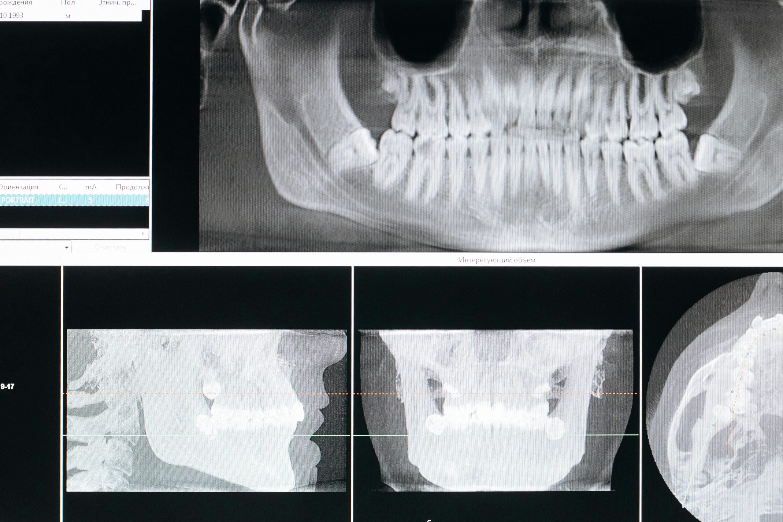
Treatment starts with careful diagnosis using clinical tests and X‑rays, and sometimes 3D imaging when needed. During treatment, a rubber dam isolates the tooth to keep saliva out, which helps prevent new bacteria from entering. The dentist then creates a small access opening, finds the canals, shapes them for cleaning, disinfects, and finally seals them to block reinfection.
Many tools support each step. Together they create accuracy and consistency:
- Rubber dam and clamps for a clean, dry working field
- Dental operating microscope for magnification and bright, focused light
- Electronic apex locator to measure canal length
- Rotary or reciprocating files to shape canals predictably
- Irrigants with ultrasonic activation to disrupt biofilm and debris
- 3D imaging (CBCT) when indicated for complex anatomy
The microscope helps reveal tiny canals, cracks, and calcifications that might be missed with loupes alone. Its coaxial light reduces shadows, which can make locating and cleaning intricate anatomy more efficient. That said, outcomes depend on the whole system: diagnosis, sterile technique, irrigation chemistry and activation, canal shaping, and the final coronal seal. To understand the microscope in endodontics value, it helps to view it as a precision aid that elevates each of these steps rather than a single fix. Many of these elements come together during comprehensive root canal treatment.
For patients, the takeaway is simple: better visibility and measurement support safer care and a higher chance of keeping your tooth. In the next section, we will look at how using a microscope may influence success and retreatment rates. Clear vision supports careful, conservative treatment.
The Role of Magnification in Dentistry
Magnification helps dentists see fine details that are hard to notice with the naked eye. By enlarging the working field and adding bright, focused light, it supports careful, conservative treatment. In endodontics and beyond, this often means more accurate diagnosis, gentler tooth preparation, and better verification of each step.
A tiny crack hides under an old filling until bright magnification reveals it. With a microscope, vision improves at the exact spot where instruments touch the tooth, so the clinician can remove less structure and still reach the target. This clarity helps locate narrow or calcified canals, check the smoothness of prepared walls, and confirm that debris is gone before sealing. In surgical endodontics, it guides precise root-end preparation and placement of repair materials.
Magnification also improves quality control. The operator can confirm margins, contacts, and contours in real time, which supports long-term fit and tissue health. That same visual feedback helps fine-tune bonded restorations, from adaptation to finishing, which can reduce roughness and staining over time. For example, enhanced visualization can aid in refining margins for custom crowns and bridges where an even edge matters.
There are practical benefits too. Better posture and a stable working distance can reduce fatigue during detailed procedures. Digital documentation through the microscope allows before-and-after images, which improves communication with patients and collaborating dentists. When judging microscope in endodontics value, the benefit shows in tasks that demand visual precision. Still, outcomes depend on the full process, including diagnosis, asepsis, irrigation, and the final restoration.
For patients, magnification can mean smaller access openings, more confident canal location, and fewer surprises later. It is one tool in a larger system aimed at keeping your tooth healthy. In the next section, we will look at how these choices may influence success and retreatment rates. Clearer vision can support more predictable care.
Benefits of Using a Microscope
A microscope lets the dentist see small details inside the tooth with bright, centered light. This clearer view helps find hidden anatomy, remove less healthy tooth, and avoid mistakes. In practical terms, it supports safer, more precise root canal care and increases the chance of keeping your natural tooth.
Picture this: a second canal hides behind a tiny dentin ledge. Under the microscope, shadows fade, the ledge is gently smoothed, and the canal is located with minimal drilling. That visibility allows conservative access, careful troughing, and smoother canal shaping. It can also reveal subtle cracks or defects earlier, which informs treatment choices before a final seal is placed.
The microscope’s coaxial light and adjustable magnification improve control at each step. During access, it helps preserve dentin while guiding instruments away from thin areas, which may reduce the risk of ledges or perforations. In calcified or curved canals, it aids in following the true path and in safely removing obstructions or old materials. Before sealing, the operator can verify that debris is cleared, irrigation has reached the target, and the chamber is clean and dry. In surgical endodontics, smaller incisions and precise root-end preparation are possible, which can limit tissue trauma and support comfortable healing.
There are workflow benefits too. A stable working distance supports steady hand control and ergonomic posture, which can improve consistency during long, detailed procedures. High-quality images and video from the microscope make it easier to document findings and explain them to patients or collaborating dentists. When weighing microscope in endodontics value, these practical gains matter most to patients. For longer visits, some patients choose mild oral sedation to stay relaxed while precise work is completed.
For patients, the result is focused care that aims to save more natural tooth and reduce surprises later. In the next section, we will look at how these benefits relate to success and retreatment rates. Clear vision guides conservative, careful care.
Comparing Microscopes and Loupes
Microscopes and loupes both make small details easier to see, but they serve different needs. Loupes are lightweight and wearable, so they are quick for exams and many routine tasks. Microscopes provide higher, adjustable magnification with bright, centered light and a stable working position, which is helpful for intricate endodontic steps. In short, loupes boost general visibility, while microscopes support very fine, precise work.
A calcified molar canal hides behind a thin dentin shelf. With loupes, the field of view is wide and mobility is excellent, which suits diagnosis, isolation, and many restorative procedures. A microscope narrows the view to the exact area of interest, adds more magnification choices, and keeps the light aligned with the line of sight to reduce shadows. Hands-free zoom and focus help the dentist refine access and follow true canal paths without extra drilling. These differences matter most when anatomy is very small, altered by age, or changed by past treatment.
Ergonomics also differ. High-power loupes can pull the neck forward, while a microscope promotes an upright posture and a consistent working distance. Team communication can improve with the microscope because images and video can be shared during and after care. Loupes remain efficient for straightforward canal identification and shaping, especially when anatomy is clear. When weighing microscope in endodontics value, consider task complexity: calcifications, suspected cracks, retreatment, and microsurgery often benefit from the microscope’s precision and documentation.
For patients, the choice influences how carefully the dentist can preserve tooth structure and avoid surprises. A microscope can support smaller access openings, more reliable canal location, and careful evaluation before sealing. Loupes still provide strong results in many cases and allow efficient, comfortable visits. Either way, vision is only one part of success, which also depends on accurate diagnosis, sterile technique, irrigation effectiveness, and a well-sealed final restoration. In the next section, we will look at how these choices may relate to success and retreatment rates. Clear vision supports careful, conservative care.
Limitations of Loupes in Endodontics
Loupes improve vision, but they have limits inside deep, narrow teeth. Their magnification is fixed, the light is not perfectly aligned with the view, and the image can dim or shadow at the back of the access cavity. As a result, very small structures, like tiny extra canals or fine cracks, can be harder to see with loupes alone.
Picture this: a faint line crosses the chamber floor beneath an old filling. With loupes, the working distance changes as the head moves, so focus and depth of field shift. At higher power, the field of view shrinks, which makes it hard to orient and follow subtle landmarks. A headlamp helps, yet it is not coaxial with the optics, so shadows and glare can hide details at the exact spot that matters.
These visual limits can affect decision making. In calcified or curved molars, loupes may require a larger access to gain line of sight, which can remove more dentin than needed. Finding a hidden canal, seeing an isthmus, or assessing the true extent of a crack is often harder without adjustable zoom and centered light. During retreatment, removing a post or bypassing a separated instrument demands precise depth cues and illumination that loupes may not provide consistently. Documentation is also limited, since still images and shared views are harder to capture through loupes, which can reduce real-time team communication.
Ergonomics matter too. High-power loupes can pull the neck forward, and small posture shifts change the angle into the tooth, which further alters light and focus. Over a long procedure, this can increase fatigue and make fine control less steady. By contrast, a stable working distance and hands-free magnification adjustments are key parts of microscope in endodontics value, especially when anatomy is small or altered by prior treatment.
For patients, this means loupes work well for many steps, but they may miss the tiniest details in complex teeth. Understanding these limits helps explain why a microscope can support more conservative access and careful canal location. Clear vision protects natural tooth structure.
Clinical Evidence Supporting Microscope Use
Research in endodontics shows that microscopes improve what clinicians can see and do. Studies report higher detection of extra canals, earlier identification of cracks, and more precise cleaning and sealing when a microscope is used. These gains are most clear in complex care, such as calcified molars, retreatment, and microsurgery.
A missed second canal in an upper molar is later found under the microscope. With bright, centered light and adjustable magnification, the operator can follow natural landmarks and uncover small canal entrances without removing excess dentin. This clearer path lowers the chance of ledges or perforations and helps instruments stay centered. Before sealing, the chamber and canal orifices can be inspected closely, supporting a cleaner interface for a tight, lasting seal.
In retreatment, magnification aids safe post removal, location of hidden anatomy, and retrieval or bypass of separated instruments. Each of these steps benefits from depth cues and shadow-free light that are difficult to achieve otherwise. In endodontic microsurgery, the microscope supports smaller osteotomies, accurate root-end preparation, and careful inspection of the resected surface for cracks or missed canals. Better visualization during root-end filling can help limit gaps and reduce tissue irritation. Together, these factors have been associated with improved healing and fewer complications compared with older techniques that lacked high magnification.
Evidence also points to consistency. When clinicians can verify each step in real time, small errors are caught earlier, so fewer surprises emerge later. Operator skill and case selection still matter, and outcomes rely on the whole process, including diagnosis, asepsis, irrigation quality, and a well-sealed final restoration. Viewed this way, the microscope strengthens critical checkpoints rather than acting as a stand-alone fix. This is the heart of microscope in endodontics value.
For patients, the message is simple: clearer vision supports conservative treatment and a better chance of keeping your tooth. Next, we will connect these findings to success and retreatment rates. Clear vision makes careful care more predictable.
Factors Influencing Treatment Outcomes
Root canal outcomes depend on many factors working together. The tooth’s condition, the level of infection, the difficulty of the anatomy, and the quality of the techniques used all matter. So do the final restoration and the patient’s overall health. No single tool guarantees success, but careful steps at each stage improve healing.
A cracked molar with a long-standing abscess needs a different plan than a fresh injury. Pre‑treatment factors shape the prognosis, including whether the pulp is alive or necrotic, the size of any lesion at the root tip, and prior dental work. Canal curvature, calcifications, and extra canals add complexity and raise the risk of missed spaces. These details guide case selection, time needed, and whether referral is wise.
Technique is the next driver. Reliable isolation with a rubber dam limits new bacteria. Accurate working length control helps clean the full canal without injuring tissues. Canal shaping should create a safe path for irrigants while conserving dentin to maintain strength. Effective irrigation, often with activation, reduces biofilm that files cannot reach. Thorough, void‑free obturation then seals the system. In this context, the microscope in endodontics value is as a precision aid that helps locate small canals, avoid errors, and verify a clean chamber before sealing.
The coronal seal strongly influences long‑term success. Prompt, well‑fitting restorations block leakage, while delayed or loose temporaries allow reinfection. Posterior teeth often benefit from cuspal coverage to reduce fracture risk. Bite forces, parafunction, and occlusion should be adjusted to protect cracks or thin walls. Patient factors also play a role. Smoking, diabetes control, dry mouth, and home care affect healing. Regular follow‑up allows early correction if symptoms return or a restoration loosens.
Operator experience and judgment tie these elements together. Choosing appropriate imaging for complex roots, recognizing when to use adjuncts, and knowing when to retreat or refer all shape outcomes. For patients, the practical steps are simple: treat infection early, complete the final restoration on time, and maintain good home care. Consistent technique, not a single device, drives healing.
Patient Experience with Advanced Technology
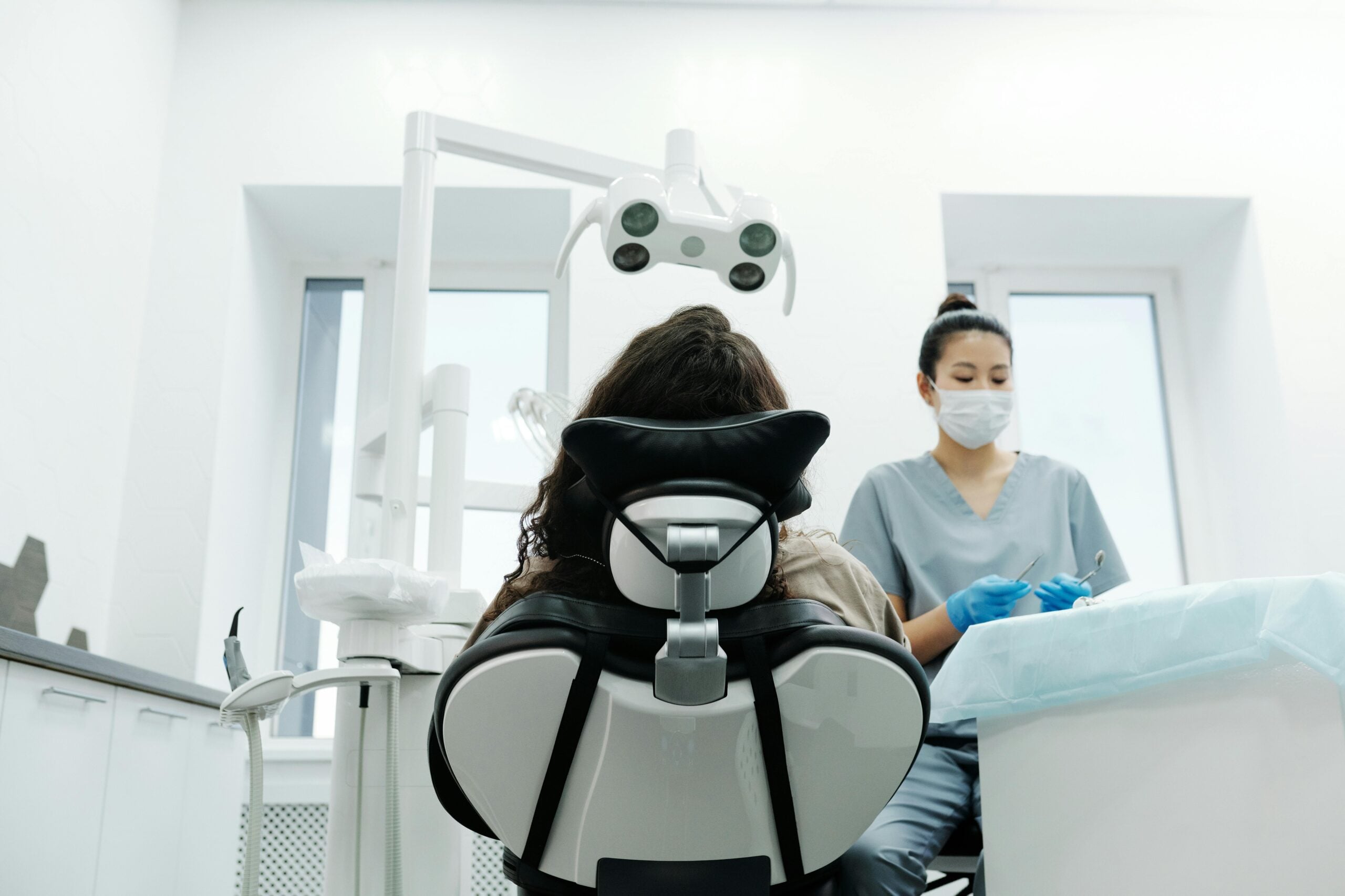
Advanced technology aims to make your visit smoother, clearer, and more predictable. With bright magnification, careful measurement tools, and modern imaging when needed, treatment can feel more focused and less disruptive. You may notice smaller access openings, steadier hands, and clearer explanations of what is happening in your tooth.
Picture this: the dentist shows you a magnified photo of a tiny canal entrance before starting. A dental operating microscope brings bright, centered light into deep spaces, so the dentist can see details without extra drilling. A rubber dam keeps the area clean and dry, which helps prevent new bacteria from entering during care. Together, these steps support gentle access and careful cleaning while preserving healthy tooth structure.
Electronic length measurement reduces guesswork, so instruments reach the intended depth without repeated adjustments. When anatomy is complex, selective 3D imaging can map curved or hidden roots before treatment begins, which reduces surprises during the procedure. Ultrasonic activation of disinfecting solutions helps disrupt debris in small fins and isthmuses that files cannot reach, supporting a thorough clean before the final seal. These tools work as a system, and this is where microscope in endodontics value becomes clear for patients.
Comfort and communication also improve. Because the microscope keeps the working distance stable, the dentist can maintain a calm, upright posture, which supports steady hand control. Digital images taken through the microscope help you see what was found and why certain choices were made. If you feel anxious or a visit may run long, some patients choose deeper sedation options after a careful health review. Your care plan is tailored to your needs and the tooth’s condition.
If you have scheduling questions, check our current hours. Clear vision, precise measurement, and a clean field help create a calmer visit and a more predictable result. The right tools work together to protect your natural tooth.
Discerning Between Equipment Options
Choosing equipment in endodontics means matching the tool to the task, not picking the fanciest device. For visibility and safety, clinicians weigh microscopes, loupes, lighting, imaging, measurement tools, and cleaning systems together. The right mix improves accuracy, preserves tooth structure, and supports a lasting seal.
Microscopes and loupes both enhance vision, but they solve different problems. Loupes are quick and mobile for exams and straightforward access. Microscopes add higher, adjustable magnification with centered light that reduces shadows deep in the tooth. During retreatment, a small post blocks the view until magnification clarifies the path. When anatomy is altered or very small, the microscope’s control over light and focus helps avoid unnecessary drilling. For simpler canals, loupes often provide sufficient clarity with good efficiency.
Imaging choices also matter. Digital radiographs guide diagnosis with low exposure, while limited field-of-view CBCT is reserved for complex roots, suspected cracks, resorption, or prior treatment that obscures landmarks. Accuracy in length control comes from combining electronic apex locators with confirming images. Cleaning decisions then follow anatomy. Ultrasonic activation can move irrigants into fins and isthmuses, while gentle agitation or manual methods may suit wider canals. Each step aims to reduce bacteria without weakening the tooth.
Instrumentation and sealing should be conservative and centered. Hand files help create a safe glide path, and modern nickel‑titanium systems shape canals while respecting curvature when selected and used carefully. Taper and size are chosen to allow irrigants to reach the target but to conserve dentin where it supports strength. For obturation, warm techniques can adapt to irregular spaces, while a single‑cone approach with a modern sealer may suit narrow or straight canals. A precise coronal restoration completes the seal. When weighing microscope in endodontics value, place it within this full system of diagnosis, measurement, disinfection, and restoration.
For patients, discerning tools means smaller openings, fewer surprises, and a stronger seal that lasts. In the next section, we will connect these choices to success and retreatment rates. The right tool at the right time protects your tooth.
Future Trends in Endodontic Technology
Future endodontic technology is moving toward safer, more precise, and more conservative care. Expect smarter imaging, digital guidance, better disinfection, and bioactive materials that protect natural tooth structure. These tools will work together, not alone, to improve diagnosis, reduce errors, and support lasting seals. The goal is clearer decisions and fewer surprises.
A calcified premolar is mapped on a focused 3D scan before access begins. Small field-of-view CBCT will be used more selectively to reveal hidden anatomy and plan conservative openings. Digital navigation and 3D-printed guides can help follow the true canal path with less drilling, especially in calcified or previously treated teeth. Early applications of artificial intelligence may aid triage, lesion detection, and case selection, supporting timely referral and targeted imaging when needed [1].
Smarter instruments are also emerging. Heat-treated nickel titanium files, adaptive motions, and torque-sensing motors aim to respect curvature while reducing file stress. Disinfection is getting a boost from more effective irrigant activation, including ultrasonics, negative pressure, and photoactivation methods that reach fins and isthmuses. Newer bioceramic sealers and hydraulic techniques can adapt to irregular spaces while keeping shapes conservative. On the biologic side, vital pulp therapy and regenerative strategies continue to develop, offering tooth-preserving options in select cases when the pulp can still heal [2].
Surgical care is evolving too. Endodontic microsurgery benefits from high magnification, micro-instruments, bioceramic repair materials, and careful root-end evaluation. These updates have been associated with improved healing and more predictable outcomes compared with older surgical methods that lacked such precision [3]. Looking ahead, these advances will extend microscope in endodontics value by linking clear visualization with digital planning, smarter motors, and bioactive materials. For patients, that means smaller openings, steadier hands, and treatment plans designed to save more of the natural tooth. Clear, coordinated tools make careful care more predictable.
Frequently Asked Questions
Here are quick answers to common questions people have about Does a Microscope Change Endo Outcomes? in Glendale, AZ.
- What is the main benefit of using a microscope in endodontics?
The main benefit of using a microscope in endodontics is enhanced visibility. This allows dentists to see finer details inside the tooth, such as hidden canals or small cracks. With better vision, endodontists can conduct more precise work, leading to safer and more effective treatment. The bright, centered light provided by the microscope reduces shadows and aids in locating intricate features without excessive tooth removal. This precision helps in preserving more of your natural tooth.
- How do microscopes and loupes differ in dental procedures?
Microscopes and loupes both improve visibility, but they serve different purposes. Loupes are lightweight and portable, suitable for quick examinations and basic dental tasks. In contrast, microscopes offer higher magnification and focused light, which are essential for complex procedures like endodontics. They provide a stable working distance and hands-free magnification adjustments, allowing for more detailed inspection and precise work, which is especially beneficial in identifying small or hidden anatomy.
- Why might a dentist choose a microscope for root canal treatment?
A dentist might choose a microscope for root canal treatment to improve precision and outcomes. The enhanced magnification and light allow for better detection of small canals, cracks, or calcifications that may not be visible with the naked eye or loupes. This technology helps ensure that all areas are thoroughly cleaned and sealed, reducing the risk of reinfection and increasing the likelihood of saving the natural tooth.
- What roles do advanced technologies play in patient comfort during dental procedures?
Advanced technologies, such as dental microscopes, enhance patient comfort by making procedures smoother and less invasive. They allow for smaller access openings in teeth, reducing the amount of drilling needed. Technologies like electronic measurers ensure precise instrument placement, and imaging techniques help avoid surprises. Together, these tools make dental visits more predictable and less stressful, ensuring that professionals work more confidently and efficiently while maintaining patient comfort.
- What limitations do loupes have compared to microscopes in endodontics?
Loupes are limited by their fixed magnification and non-centered light, which can produce shadows and a dimmer image. These factors make it challenging to view deep, narrow areas inside a tooth, where vital tiny structures like extra canals or fine cracks might be present. A microscope offers adjustable magnification and coaxial lighting, ensuring a clearer and more detailed view, which is crucial for preventing treatment errors in complex cases.
References
- [1] On the Horizon: What’s Next for Endodontics. (2025) — PubMed:41106913 / DOI: 10.1016/j.cden.2025.05.007
- [2] Advances in clinical and translational research in endodontics: A comprehensive overview. (2025) — PubMed:41172500 / DOI: 10.1590/0103-644020256723
- [3] Optimising Outcomes in Endodontic Microsurgery: Evidence, Uncertainties and Future Directions. (2026) — PubMed:41555382 / DOI: 10.1111/iej.70100